Download
1 / 14
170 likes | 818 Views
Raman spectroscopy. Mr/ Abd El mo’ez Ahmed Mohammed “ 3th group ”. “ Neutron physics and application in material science and nuclear application ” physics department Faculty of Science Sohag University Egypt. Physics Department. Faculty of Science. JINR’s group.

E N D
Mr/ Abd El mo’ez Ahmed Mohammed“3th group” “Neutron physics and application in material scienceand nuclear application”physics departmentFaculty of ScienceSohag University Egypt Physics Department Faculty of Science
JINR’s group Supervisor : prof . S.I. Tutunnikov. Technician : Mr. Kovalev Yura.
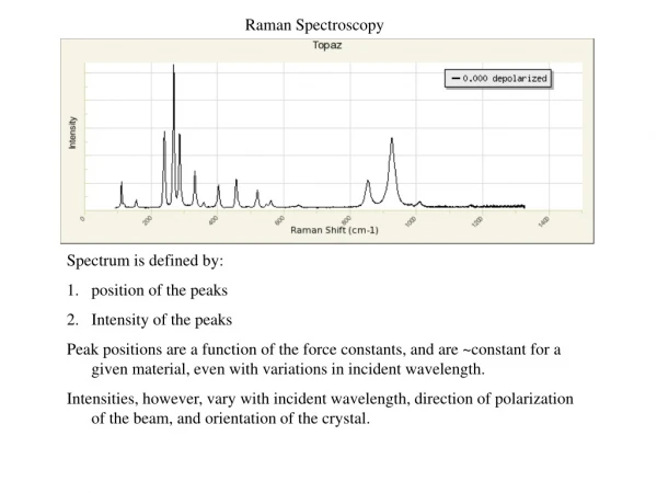
Why we use Raman spectroscopy? Raman spectroscopy is used to determine the molecular motions, especially the vibrational one .
Now what is the application? We can use Raman spectroscopy in:- Tissue characterization. Tissue Imaging. Live Cell Imaging.
Live Cell Imaging All the sexiness, without the dyes!!
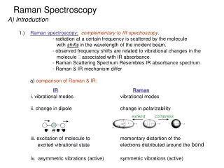
difference in energy hn h(n (-+) n1) 3 2 1 0 S1 3 2 1 0 S0 hn Inelastic Scattering Virtual Level • Energy transferred from incident light to molecular vibrations Energy Rayleigh Raman (inelastic) (elastic) Scattering Scattering
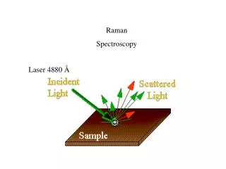
Raman spectrometer’s mechanism. The laser beam falls on the filter and then it passes to the objective and collides with the Sample. The light will be scattered in all directions and will be reflected to the objective again. After the reflected light reaches the objective it falls down on the spectrometer system and finally to the CCD or PMT detectors.
Acknowledgement Finally I’d like to thank 1- Mr/ Kovalev Yura. 2- Mr/ Valantine. for their help and efforts.