Download
1 / 26
260 likes | 480 Views
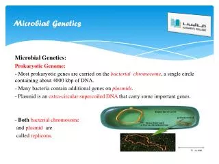
Microbial groups. CE 421/521 Lecture September 14, 2006 Vaccari et al., Chapter 10. Microbes. Microorganisms – broad category of organisms too small to be seen with the naked eye Integral part of every ecosystem Roughly 10 6 to 10 9 per gram of soil, biofilm or sludge sample.

E N D
Microbial groups CE 421/521 Lecture September 14, 2006 Vaccari et al., Chapter 10
Microbes • Microorganisms – broad category of organisms too small to be seen with the naked eye • Integral part of every ecosystem • Roughly 106 to 109 per gram of soil, biofilm or sludge sample
Microbial groups • Prokaryotes • Bacteria (Including blue green algae) • Archae – (sometimes archaebacteria) • classified during the 1970’s by Carl Woese and George Fox • Don’t fit neatly into prokaryotic or eukaryotic class due to their difference in 16S rRNA - separate kingdom? • Includes methanogens and halophiles • Viruses – Dimitri Ivanovsky (1893) filtered sap through ceramic filters designed to remove bacteria – still resulted in tobacco mosaic virus • Eukaryotes
Classification of microorganisms • Energy source: • Chemotrophs – energy from chemical substances • Organotrophs – energy from organic compounds • Lithotrophs – energy from an inorganic compound • Phototrophs – energy from sunlight
Classification of microorganisms • Carbon source: • Heterotrophs – carbon from organic compounds • Autotrophs – carbon from inorganic compounds Can have mixed classifications: e.g. chemoorganoheterotroph (example E. Coli) chemolithoautotroph (example nitrobacter)
Classification of microorganisms • Environmental preferences: • TEA (anaerobic, aerobic, anoxic) • Temperature • Psychrophiles • Mesophiles • Thermophiles • pH • Neutrophiles (5-9) • Acidophiles (< 5) • Alkaliphiles (> 9) • Extremophiles – can grow at extreme temperatures or osmotic pressures (e.g., halophiles)
Microbial Taxonomy • Morphology: form and visible structure • Biocehmcial activities • Phenotype – representing observable characteristics • Genotype • Characterized by DNA or RNA • Phylogeny – based on genetic similarities
Taxonomy - What is a prokaryotic species? • Difficulty in that genetic exchange occurs between species not necessarily closely related • Strain • have a recent parent cell • Share genetic properties with minor exceptions • Species • Share at least 70% of DNA homology – similarity in DNA sequence • Or have rRNA similarity of 97% or greater • Genus • Share at least 20% of their DNA homology • Or have rRNA similarity of 93-95%
Nomencalture • E_______________ (e.g., aquaticus, marina, coli) • H______ (e.g., bovus, avium) • Environmental c__________ (e.g., thermophilus, halophilus) • S_______ (e.g., ovalis, longum, spaericus) • C_______ (e.g., aureus, niger) • S_______________ (e.g., denitrificans, avium) • P____________ (e.g., methanobacterium, cerevisiae) • D_____________ (e.g., typhi, botulinum, pneumoniae) • P_____________ (e.g., winogradskii, burkholderia)
Prokaryotes - shape • cocci (spherical, e.g., Streptococcus) • bacilli (rod shapes, e.g., Bacillus subtilis) • spirilla (spiral, e.g., Spirillum volutans) • filamentous
Prokaryotes - shape • Unusual • s___________ bacteria - filamentous, surrounded by a sheath • s___________ bacteria - aerobic, gram negative, at end of stalk is a “holdfast” allows it to attach to surfaces • b___________ bacteria, multiply by budding, bud grows flagellum, settles on new surface and buds again • g___________ bacteria, filamentous, gram-negative, “glide” along solids surfaces, Beggiatoa and Thiothrix: oxidize H2S to S0
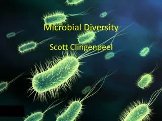
Prokaryotes - shape • Bdellovibrio - s________ (0.2-0.3μ) flagellated bacteria that prey on gram-negative bacteria • Actinomycetes- gram-positive, f_________________, have branching filaments similar to fungi - Streptomyces and Nocardia • Cyanobacteria - b____________-g___________ algae, procaryotes, contain chlorophyl a, have characteristic blue-green color, contain gas vacuoles that enable them to float to maximize photosynthesis, responsible for algal blooms, some are toxic
http://plpnemweb.ucdavis.edu/nemaplex/images/actinomycete.jpghttp://plpnemweb.ucdavis.edu/nemaplex/images/actinomycete.jpg Actinomycetes http://www.visualsunlimited.com/images/watermarked/188/188934.jpg Bdellovibrio
http://www.cbcis.wustl.edu/images/27-11x1-Cyanobacteria.jpg Cyanobacteria
Fungi • e_________________, produce long filaments called hyphae containing c___________ • heterotrophs, use o________________ compounds for carbon and energy • found during n______________ limitations, low D.O., low pH conditions • important in the cycling of organics – degradation of plant polymers cellulose and lignin • primarily aerobic (except for fermentative yeast)
Algae • most are u___________________, floating, phytoplankton • some are f____________________ • most are p____________________ • all contain chl____________________ a, some b and c • found in o____________________ ponds, polishing ponds, aerobic lagoons
Protozoa • Unicellular • Heterotrophs • Classification • sarcodina (amoebae) • mastigophora (flagellates) • ciliophora (ciliates) • sporozoa
Viruses • small c________________ particles (not procaryotes or eucaryotes) are they alive? • replication occurs in h_______________ • Structure • c___________ of nucleic acid (could be double or single stranded, DNA or RNA) surrounded by protein coat (capsid) • main shapes • h_________________ • p_________________ • c_________________
Virus Replication • Ad______________________ - virus adsorbs to specific receptors, receptors can be polysaccharides, proteins, or lipoproteins • En______________________ - various particle or nucleic acid material enters cell • Ec______________________ - capsid is stripped away, releasing genetic material • Mu______________________ - viral nucleic acids are replicated using machinery of host cell • Ma______________________ - protein coat is synthesized and combined with nucleic acid to form nucleocapsid • Re_______________________ of mature virions - host cell ruptures release active viruses
Virus Detection and Enumeration • animal i___________________ - newborn mice injected with inoculum and observed for signs of disease • t_____________ cultures - viruses quantified by measuring effect on host cell lines forming a monolayer on glass or plastic assay bottles, effect is measure by • p________________ assay - virus is placed on surface of host cell monolayer, virus replication leads to localized area of cell destruction called plaques • s____________ dilution endpoint - virus suspension is diluted serially and the highest dilution (smallest amount of virus) that causes a cytopathic effect in 50% of samples is reported as the tissue culture infectious dose (TCID50) • most p_____________ number - serial dilutions placed in tubes or microwells with host cells, positive tubes are recorded and MPN value computed from standardized MPN table.
MPN • Uses serial dilutions and statistical probabilities for the most likely number of organisms giving a positive response
Example: Take 1 mL of sample and add to 1 L of water then perform the following serial dilutions:10 mL, 1 mL, and 0.1 mL and incubate with substrate. If we get 5 positive tubes in the first dilution, 4 positive tubes in the second dilution, and 1 in the last dilution, what is the MPN of the sample? Solution: from the following table we can see that the as diluted MPN is 170. Since we had a 1000 fold dilution to start with, the resulting MPN is 170,000 organisms per 100 mL