Download

1 / 76
790 likes | 924 Views
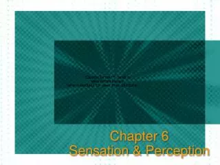
0. Chapter 2: The Beginnings of Perception. Figure 2-1 p22. Light: the Stimulus for Vision. Electromagnetic spectrum Energy is described by wavelength. Spectrum ranges from short wavelength gamma rays to long wavelength radio waves.

E N D
0 Chapter 2: The Beginnings of Perception
Light: the Stimulus for Vision • Electromagnetic spectrum • Energy is described by wavelength. • Spectrum ranges from short wavelength gamma rays to long wavelength radio waves. • Visible spectrum for humans ranges from 400 to 700 nanometers. • Most perceived light is reflected light
The Eye • The eye contains receptors for vision • Light enters the eye through the pupil and is focused by the cornea and lens to a sharp image on the retina. • Rods and cones are the visual receptors in the retina that contain visual pigment. • The optic nerve carries information from the retina toward the brain.
Light is Focused by the Eye • The cornea, which is fixed, accounts for about 80% of focusing. • The lens, which adjusts shape for object distance, accounts for the other 20%. • Accommodation results when ciliary muscles are tightened which causes the lens to thicken. • Light rays pass through the lens more sharply and focus near objects on retina.
Loss of Accommodation With Increasing Age • The near point occurs when the lens can no longer adjust for close objects. • Presbyopia - “old eye” • Distance of near point increases • Due to hardening of lens and weakening of ciliary muscles • Corrective lenses are needed for close activities, such as reading
Myopia • Myopia or nearsightedness - Inability to see distant objects clearly • Image is focused in front of retina • Caused by • Refractive myopia - cornea or lens bends too much light • Axial myopia - eyeball is too long
Focusing Images on Retina - continued • Solutions for myopia • Move stimulus closer until light is focused on the retina • Distance when light becomes focused is called the far point. • Corrective lenses can also be used. • LASIK surgery can also be successful.
Hyperopia • Hyperopia or farsightedness - inability to see nearby objects clearly • Focus point is behind the retina. • Usually caused by an eyeball that is too short • Constant accommodation for nearby objects can lead to eyestrain and headaches.
Transforming of Light Energy Into Electrical Energy • Receptors have outer segments, which contain: • Visual pigment molecules, which have two components: • Opsin - a large protein • Retinal - a light sensitive molecule • Visual transduction occurs when the retinal absorbs one photon. • Retinal changes it shape, called isomerization.
Transforming of Light Energy Into Electrical Energy - continued • Current research in physiology and chemistry shows that isomerization triggers an enzyme cascade. • Enzymes facilitate chemical reactions. • A cascade means that a single reaction leads to increasing numbers of chemical reactions. • This is how isomerizing one pigment leads to the activation of a rod receptor.
Adapting to the Dark • Dark adaption is the process of increasing sensitivity in the dark
Distribution of Rods and Cones • Differences between rods and cones • Shape • Rods - large and cylindrical • Cones - small and tapered • Distribution on retina • Fovea consists solely of cones. • Peripheral retina has both rods and cones. • More rods than cones in periphery.
Distribution of Rods and Cones - continued • Macular degeneration • Fovea and small surrounding area are destroyed • Creates a “blind spot” on retina • Most common in older individuals • Retinitis pigmentosa • Genetic disease • Rods are destroyed first • Foveal cones can also be attacked • Severe cases result in complete blindness
Distribution of Rods and Cones - continued • Number - about 120 million rods and 6 million cones • Blind spot - place where optic nerve leaves the eye • We don’t see it because: • one eye covers the blind spot of the other. • it is located at edge of the visual field. • the brain “fills in” the spot.
Measuring the Dark Adaptation Curve • Three separate experiments are used. • Method used in all three experiments: • Observer is light adapted • Light is turned off • Once the observer is dark adapted, she adjusts the intensity of a test light until she can just see it.
Measuring the Dark Adaptation Curve - continued • Experiment for rods and cones: • Observer looks at fixation point but pays attention to a test light to the side. • Results show a dark adaptation curve: • Sensitivity increases in two stages. • Stage one takes place for three to four minutes. • Then sensitivity levels off for seven to ten minutes - the rod-cone break. • Stage two shows increased sensitivity for another 20 to 30 minutes.
Measuring the Dark Adaptation Curve - continued • Experiment for cone adaptation • Test light only stimulates cones. • Results show that sensitivity increases for three to four minutes and then levels off. • Experiment for rod adaptation • Must use a rod monochromat • Results show that sensitivity increases for about 25 minutes and then levels off.
Visual Pigment Regeneration • Process needed for transduction: • Retinal molecule changes shape • Opsin molecule separates • The retina shows pigment bleaching. • Retinal and opsin must recombine to respond to light. • Cone pigment regenerates in six minutes. • Rod pigment takes over 30 minutes to regenerate.
Spectral Sensitivity • Sensitivity of rods and cones to different parts of the visual spectrum • Use monochromatic light to determine threshold at different wavelengths • Threshold for light is lowest in the middle of the spectrum • 1/threshold = sensitivity, which produces the spectral sensitivity curve
Spectral Sensitivity - continued • Rod spectral sensitivity shows: • more sensitive to short-wavelength light. • most sensitivity at 500 nm. • Cone spectral sensitivity shows: • most sensitivity at 560 nm. • Purkinje shift - enhanced sensitivity to short wavelengths during dark adaptation when the shift from cone to rod vision occurs
Spectral Sensitivity - continued • Difference in spectral sensitivity is due to absorption spectra of visual pigments • Rod pigment absorbs best at 500 nm. • Cone pigments absorb best at 419nm, 531nm, and 558nm • Absorption of all cones equals the peak of 560nm in the spectral sensitivity curve
Electrical Signals in Neurons • Key components of neurons: • Cell body • Dendrites • Axon or nerve fiber • Sensory receptors - specialized neurons that respond to specific kinds of energy
Recording Electrical Signals in Neurons • Small electrodes are used to record from single neurons. • Recording electrode is inside the nerve fiber. • Reference electrode is outside the fiber. • Difference in charge between them is -70 mV • This negative charge of the neuron relative to its surroundings is the resting potential.