Download
1 / 59
590 likes | 809 Views
When should you wriggle your TOE’s in Theatre?. Andrew Ronald Consultant Anaesthetist Aberdeen Royal Infirmary andrew.ronald@nhs.net. Is there a role for Transoesophageal Echocardiography (TOE) in Non-Cardiac Surgery?. TOE in Non-Cardiac Surgery. Introduction to TOE

E N D
When should you wriggle your TOE’s in Theatre? Andrew Ronald Consultant Anaesthetist Aberdeen Royal Infirmary andrew.ronald@nhs.net
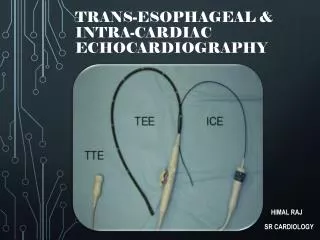
Is there a role for Transoesophageal Echocardiography (TOE) in Non-Cardiac Surgery?
TOE in Non-Cardiac Surgery • Introduction to TOE • What does TOE tell us that other monitors don’t • Indications and Contraindications • Can we identify Non-Cardiac Surgical patients who might benefit from TOE, and is there any evidence base to support that assertion? • Are there disadvantages to TOE? • When do I think we should consider using TOE
A Brief History of TOE • 1953/54: Edler & Hertz - “Ultrasound cardiography” • 1955/56: Yoshida - Doppler ultrasound to detect cardiac motion • 1971: 2D Echocardiography introduced • 1974-76: Development of Transoesophageal Echocardiography (TOE) • early 2000’s: Development of 3D-Echo • 2006-2007: Introduction of real-time 3D TOE into clinical practice
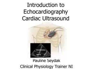
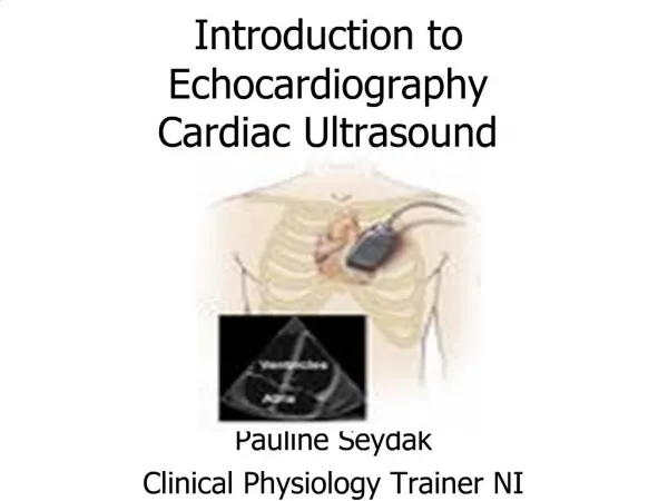
Echo is very high frequency sound (2-10 MHz) follow physical laws of reflection and refraction and can be focused and orientated like beams reflected by dense materials and attenuated at depth propagates freely through liquids but poorly through air return of waveform signal dependant on structures it hits timed firing of multiple “phased array” of piezo-electric crystals can create a “scanning front” across area of interest Physics of TOE f=frequency cycles/sec; Hz) =wavelength c=speed of sound in tissues and blood ~ 1540m/sec f = 1/T C = f Tissue penetration
Physics of TOE • “US transducers on gastroscope body” • imaging from oesophagus • insertion / withdrawl of probe • anteflexion / retroflexion / lateral flexion • rotation of US beam axis • series of standard views recommended by ASA / SCA
Modes of Echocardiography • 2D echo • M-mode • Doppler • 3D Echo
2-Dimensional Echocardiography • Conventional anatomical tomography of structures within field • Real-time 2-D assessment of myocardial structure / function • Frequency, beam width, pulse length & transducer radius affect resolution • Freeze / cine-loop enables off-line analysis
M-mode Echocardiography • Charts movements of structures along an interrogation line against time • Useful for rapidly moving structures • Useful for accurate measurements of cardiac dimensions
Doppler Echocardiography • Doppler echocardiography • “…..apparent change in received frequency due to relative motion between a sound source and sound receiver….” • Fd=2FtVCos/C • Ft= transmitted Doppler freq • V= blood flow • Cos= cosine of angle between blood flow and US beam (Cos 0=1; Cos 90=0) • C= speed of sound in tissue • P=4V2
3-Dimensional Echocardiography • Until recently unavailable in “real-time” • Involves acquisition of large amounts of data, only a fraction of which is actually used for 3DE reconstruction • Allows visualisation of structures from multiple viewpoints
TOE in Non-Cardiac Surgery What does TOE tell us that other monitors don’t?
What does TOE tell us? • Dynamic Real-Time Anatomy, Physiology and Pathology • Dynamic Cardiac Function • You can see the heart and the proximal great vessels • Changes in wall motion precede all other signs of ischaemia • Monitor response to interventions directly • Direct measurement / monitoring of systolic & diastolic function • FAC, FS, EF, SV, CO • Dynamic Valvular function • Normal / Abnormal • Tailor management to abnormality • Monitor response to interventions directly
What does TOE tell us? “……..a picture is worth a thousand words……..”
TOE in Non-Cardiac Surgery Indications & Contraindications
Contraindications to TOE • Absolute Contraindications • Cardiac practice - probably no absolute contraindications • Non-cardiac practice - gastro-oesophageal surgery • Relative Contraindications • varices • oesophageal tumour • UGI bleeding • Use with care • dysphagia • hiatus hernia • anticoagulation
Category I indications • supported by strongest evidence / expert opinion • TOE frequently useful in improving outcome • Category II indications • supported by weaker evidence / expert opinion • TOE may be useful in improving outcome • Category III indications • little current evidence / expert opinion • TOE infrequently useful in improving outcome Do any of the general indications identified for Cardiac Surgery extrapolate to Non-Cardiac surgery?
What is a Category I indication? “Evaluation of acute, persistent, life threatening haemodynamic function in patients in whom ventricular function and its determinants are uncertain and who have not responded to treatment” TOEfrequently useful in improving outcome
What is a Category II indication? “Perioperative use in patients at increased risk of myocardial ischaemia / infarction, haemodynamic disturbance or cardiac trauma” TOE may be useful in improving outcomes
Why is TOE useful in patients with risk of myocardial ischaemia / infarction • Regional wall motion abnormalities precede ECG changes so myocardial ischaemia recognised early • Indirect evidence that early Rx of myocardial ischaemia and MI improves outcomes
Why is TOE useful in patients with risk of haemodynamic disturbance? • Quantitative assessment of ventricular dimensions enhances conventional monitoring • Better assessment of pre-load (EDV) than PAC
Why is TOE useful in patients with(Cardiac) Trauma? • (Usefulness of TOE in diagnosing Thoracic Aortic injuries well recognised) • Shortcomings of other methods of assessing adequacy of LV filling well recognised • Facilitates direct assessment of myocardial filling / emptying and “obstructions” to these e.g. tamponade • (Facilitates direct assessment of cardiac valve competency and myocardial thickening)
Can we identify patients from a non-cardiac surgical population who in theory at least might benefit from TOE?
Category I Indications • Probably a bit of a “no-brainer” • “Evaluation of acute, persistent, life threatening haemodynamic function in which ventricular function and its determinants are uncertain and have not responded to treatment” • ACC / AHA / ASE 2003 Guideline Update on the Clinical Application of Echocardiography • “Emergency use to determine the cause of an acute persistent and life-threatening haemodynamic disturbance” • Recommendation in 2007 ACC / AHA Guideline on Perioperative Cardiovascular Evaluation and Care for Non-Cardiac Surgery (Circulation 2007 ; 116: e418-e500)
Category II indications • Increased risk of myocardial ischaemia / infarction • Increased risk of haemodynamic disturbance • How do we identify these patients most at risk who might benefit? • More more difficult!
Revised Cardiac Risk IndexLee et al Circulation 1999; 100: 1043-1049 • Major risk factors • High-risk type of surgery • History of Ischaemic heart disease • History of Congestive heart failure • History of Cerebrovascular disease • Preoperative Rx with Insulin • Preoperative Serum Creatinine > 2mg/dl (180 mol/l)
Cardiac Risk Indexes • May give us some sort of starting point - but what is it? • Do we TOE everyone with >3 risk factors? • Clearly impractical – large numbers • Tells us nothing on changing outcomes • The question remains, at what level of risk do we consider perioperative TOE? • Does it influence our outcomes in non-cardiac surgery?
Intraoperative transoesophageal echocardiography during non-cardiac surgery Suriani RJ et al. J Cardioth & Vasc Anesth 1998; 12: 274-280 Perioperative use of transoesophageal Echocardiography by Anesthesiologists: impact in non-cardiac surgery and in the intensive care unit Denault AY et al. Can J Anesth 2002; 49: 287-293 Therapeutic impact of intra-operative transoesophageal echocardiography during non-cardiac surgery Hofer CK et al. Anaesthesia 2004; 58: 3-9 Impact of intraoperative transoesophageal echocardiography during non-cardiac surgery Schulmeyer MCC et al. J Cardioth & Vasc Anesth 2006; 20: 768-771 Use of transoesophageal echocardiography during cardiac arrest in patients undergoing elective non-cardiac surgery Lin T ey al. Br J Anaesth 2005; 96: 167-70
What do they tell us? • All show evidence that TOE can be used to guide / change therapy in the perioperative period • Studies suggest TOE especially useful in the presence of Category I and less so in Category II indications • TOE use can affect not only drug and fluid management but can also influence surgical management
What do they tell us? • TOE would appear to be particularly useful in management of patients with pre-op RWMA & history of LVF, or Rt heart failure • TOE is particularly useful in picking up “new” LV, RV or valvular dysfunction • TOE may be useful in the non-cardiac surgery peri-arrest situation • TOE may be used as a PAC substitute
Why we should be cautious • Level of evidence - all studies reported are essentially retrospective or prospective Cohort studies. PRCT’s would give us a better level of evidence – but clearly near impossible to perform in this study group • There may be an “inherent bias” in retrospective review of the “usefullness of TOE” by TOE operators in certain studies. • Mainly North American studies • Inclusion criteria based mostly around old Category I and Category II indications – really need more solid indications, criteria and “level of risk” for intervention.
Why we should be cautious • Some studies very protocol driven • How does TOE compare to other monitoring modalities? There is a distinct lack of comparative studies. • TOE may change perioperative management, but does it change long-term outcome? • Although assumption that improved intra-operative management means better long-term outcomes probably valid, none of the non-cardiac studies actually demonstrate this
Are there disadvantages to TOE? • Expensive! • TOE machine ~ £100K • Probes ~ £20K • Patient safety • Consent issues • Major morbidity • Major complication rate 0-0.5% • Mortality 0.004% • Minor morbidity • Common • Who does it? • Cardiologist • Echocardiographer / Radiologist • Anaesthetist
Are there disadvantages to TOE? • Training issues • Intubation, image acquisition, handling & interpretation • Understanding of the implications of the perioperative setting • “Basic” scanning vs “full” scanning • Skills retention • British Society of Echocardiography very clear as regards numbers, process and time course for accreditation • How many do you need to perform to retain your skills? • Is there any proven cost benefit? • For monitoring – not currently • Lack of studies showing improvement in outcome
TOE in Non-Cardiac Surgery When do I think we should consider using Perioperative TOE?
My Indications for Perioperative TOE • Diagnostic tool • Not primarily. Diagnosis should be made prior to surgery • Chance findings / diagnostic changes • Monitoring tool • Ultimate Cardiac monitor • Subjective vs Objective - “Look” vs “Measure” • use all available modes of Echo • Monitor responses to therapy • Other “monitoring”
My Category I Indications for perioperative TOE • Emergency use to determine the cause of an acute persistent and life-threatening haemodynamic disturbance
69 year old male • General surgery - splenectomy for myelofibrosis • PMH incl previous MI (mild LV impairment); CVA • Cardiac arrest day 4 on ward • Resuscitated – returned to theatre for laparoscopy / laparotomy - ? Intra-abdominal haemorrhage - negative • Low BP despite inotropes • High CVP ? PE • On-table TOE requested
No PE Probable diagnosis – cardiac arrest 2° to myocardial ischaemia
My possible Category II Indications for perioperative TOE • Increased risk of haemodynamic instability • Patients with significantly impaired cardiac function undergoing high-risk surgery • A combination of both I cannot say what level of “risk” justifies TOE monitoring – “Risk Indexes” may in future provide a “starting point”
Increased risk of haemodynamic instability severe cardiac disease aortic surgery extensive cancer surgery patients with HOCM risk of tumour or thrombus embolisation to heart with RVOT obstruction thoracic trauma Patients with significantly impaired cardiac function undergoing high-risk surgery severe cardiac failure severe LV dysfunction HOCM cardiomyopathy severe valvular disease
28 year old female • Gynaecological / General surgery • Presented with abdominal pain / vomiting • USS - ? Thrombus in IVC • CT – mass extending from iliac veins to IVC • Diagnosis – likely pelvic malignancy (? Uterine) extending up IVC • PMH – Myomectomy for uterine fibroid 3 yrs previously • TOE requested to monitor thrombus / tumour in IVC and monitor for possible embolisation
Removal of IVC tumour / thrombus No obvious perioperative tumour / thrombus embolism to Rt heart
65 year old female • General & urology (& Cardiac) • Renal tumour with tumour / extension in IVC and RA • TOE requested to monitor thrombus / tumour in IVC and monitor for possible embolisation