Download

1 / 36
360 likes | 500 Views
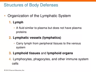
Structures of Body Defenses. Figure 20–6b Lymphoid Nodules. Structures of Body Defenses. Distribution of Lymphoid Nodules Lymph nodes Spleen Respiratory tract (tonsils) Along digestive and urinary tracts. Structures of Body Defenses. Mucosa-Associated Lymphoid Tissue (MALT)

E N D
Structures of Body Defenses Figure 20–6b Lymphoid Nodules.
Structures of Body Defenses • Distribution of Lymphoid Nodules • Lymph nodes • Spleen • Respiratory tract (tonsils) • Along digestive and urinary tracts
Structures of Body Defenses • Mucosa-Associated Lymphoid Tissue (MALT) • Lymphoid tissues associated with the digestive system • Aggregated lymphoid nodules • Clustered deep to intestinal epithelial lining • Appendix (or vermiform appendix) • Contains a mass of fused lymphoid nodules
Structures of Body Defenses • The Five Tonsils • In wall of pharynx • Left and right palatine tonsils • Pharyngeal tonsil (adenoid) • Two lingual tonsils
Structures of Body Defenses • Lymphoid Organs • Lymph nodes • Thymus • Spleen • Are separated from surrounding tissues by a fibrous connective tissue capsule
Structures of Body Defenses • Lymph Nodes • Trabeculae • Bundles of collagen fibers • Extend from capsule into interior of lymph node • Hilum • A shallow indentation where blood vessels and nerves reach the lymph node
Structures of Body Defenses • Lymph Nodes • Afferent lymphatic vessels • Carry lymph: • from peripheral tissues to lymph node • Efferent lymphatic vessels • Leave lymph node at hilum • Carry lymph to venous circulation
Structures of Body Defenses Figure 20–7 The Structure of a Lymph Node.
Structures of Body Defenses • Lymph from Afferent Lymphatics • Flows through lymph node in a network of sinuses • From subcapsular space: contains macrophages and dendritic cells • Through outer cortex: contains B cells within germinal centers • Through deep cortex: dominated by T cells • Through the core (medulla): contains B cells and plasma cells, organized into medullary cords • Finally, into hilum and efferent lymphatics
Structures of Body Defenses • Lymph Node • A filter • Purifies lymph before return to venous circulation • Removes • Debris • Pathogens • 99% of antigens
Structures of Body Defenses • Antigen Presentation • First step in immune response • Extracted antigens are “presented” to lymphocytes • Or attached to dendritic cells to stimulate lymphocytes
Structures of Body Defenses • Lymphoid Functions • Lymphoid tissues and lymph nodes • Distributed to monitor peripheral infections • Respond before infections reach vital organs of trunk
Structures of Body Defenses • Lymph Nodes of Gut, Trachea, Lungs, and Thoracic Duct • Protect against pathogens in digestive and respiratory systems
Structures of Body Defenses • Lymph Nodes (Glands) • Large lymph nodes at groin and base of neck • Swell in response to inflammation • Lymphadenopathy • Chronic or excessive enlargement of lymph nodes may indicate infections, endocrine disorders, or cancer
Structures of Body Defenses • The Thymus • Located in mediastinum • Atrophies after puberty • Diminishing effectiveness of immune system • Divisions of the Thymus • Thymus is divided into two thymic lobes • Septa divide lobes into smaller lobules
Structures of Body Defenses • A Thymic Lobule • Contains a dense outer cortex and a pale central medulla • Lymphocytes • Divide in the cortex • T cells migrate into medulla • Mature T cells leave thymus by medullary blood vessels
Structures of Body Defenses • Reticular Epithelial Cells in the Cortex • Surround lymphocytes in cortex • Maintain blood–thymus barrier • Secrete thymic hormones that stimulate • Stem cell divisions • T cell differentiation
Structures of Body Defenses • Reticular Epithelial Cells in the Medulla • Form concentric layers known as thymic (Hassall) corpuscles • The medulla has no blood–thymus barrier • T cells can enter or leave bloodstream • Thymus Hormones • Thymosin, an extract from the thymus that promotes development of lymphocytes
Structures of Body Defenses Figure 20–8 The Thymus.
Structures of Body Defenses Figure 20–8 The Thymus.
Structures of Body Defenses Figure 20–8 The Thymus.
Structures of Body Defenses Figure 20–8 The Thymus.
Structures of Body Defenses • Three Functions of the Spleen • Removal of abnormal blood cells and other blood components by phagocytosis • Storage of iron recycled from red blood cells • Initiation of immune responses by B cells and T cells • In response to antigens in circulating blood
Structures of Body Defenses • Structure of the Spleen • Attached to stomach by gastrosplenic ligament • Contacts diaphragm and left kidney • Splenic veins, arteries, and lymphatic vessels • Communicate with spleen at hilum
Structures of Body Defenses • Structure of the Spleen • Inside fibrous capsule • Red pulp: contains many red blood cells • White pulp: resembles lymphoid nodules
Structures of Body Defenses • Trabecular Arteries • Branch and radiate toward capsule • Finer branches surrounded by white pulp • Capillaries discharge red blood cells into red pulp • Red Pulp • Contains elements of circulating blood • Plus fixed and free macrophages
Structures of Body Defenses • Splenic Circulation • Blood passes through • Network of reticular fibers • Then enters large sinusoids (lined by macrophages) • Which empty into trabecular veins
Structures of Body Defenses Figure 20–9 The Spleen.
Structures of Body Defenses Figure 20–9 The Spleen.
Structures of Body Defenses • Spleen Function • Phagocytes and other lymphocytes in spleen • Identify and attack damaged and infected cells • In circulating blood
Structures of Body Defenses • Body defenses provide resistance to fight infection, illness, and disease • Two categories of defenses • Nonspecific defenses • Specific defenses • Nonspecific and specific defenses operate together to provide resistance to infection and disease
Structures of Body Defenses • Nonspecific Defenses • Always work the same way • Against any type of invading agent • Specific Defenses • Protect against specific pathogens • Depend on activities of lymphocytes • Specific resistance (immunity) • Develops after exposure to environmental hazards
Nonspecific Defenses • Seven major categories of nonspecific defenses • Physical barriers • Phagocytes • Immunological surveillance • Interferons • Complement • Inflammatory response • Fever
Nonspecific Defenses • Physical Barriers • Keep hazardous materials outside the body • Phagocytes • Attack and remove dangerous microorganisms • Immunological Surveillance • Constantly monitors normal tissues • With natural killer cells (NK cells)
Nonspecific Defenses • Interferons • Chemical messengers that trigger production of antiviral proteins in normal cells • Antiviral proteins • Do not kill viruses • Block replication in cell • Complement (C) Proteins • Form the complement system • Complement action of antibodies