Download
1 / 8
80 likes | 226 Views
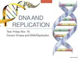
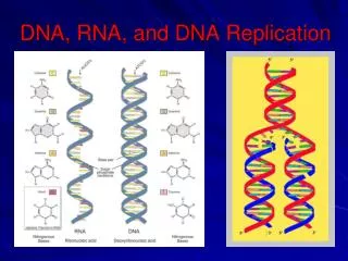
DNA and Replication. Fig. 5-27. Making a nucleotide. 5 end. Nitrogenous bases Pyrimidines. 5 C. 3 C. Nucleoside. Nitrogenous base. Thymine (T, in DNA). Uracil (U, in RNA). Cytosine (C). Purines. Phosphate group. Sugar (pentose). 5 C. Adenine (A). Guanine (G).

E N D
Fig. 5-27 Making a nucleotide 5 end Nitrogenous bases Pyrimidines 5C 3C Nucleoside Nitrogenous base Thymine (T, in DNA) Uracil (U, in RNA) Cytosine (C) Purines Phosphate group Sugar (pentose) 5C Adenine (A) Guanine (G) (b) Nucleotide 3C Sugars 3 end (a) Polynucleotide, or nucleic acid Nitrogenous base connected to 1’ carbon of sugar – nucleoside Ribose (in RNA) Deoxyribose (in DNA) Phosphate group added to 5’ carbon of sugar - nucleotide (c) Nucleoside components
5’ C (free) DNA is a linear polymer of nucleotide subunits joined together by phosphodiester bonds - covalent bonds between phosphate group at 5’ carbon and 3’ carbon of next nucleotide – uses oxygens as bridges. Chain of nucleotides has alternating sugar and phosphate components, called the “sugar-phosphate backbone.” Nitrogenous bases stick off backbone at regular intervals. Note that any linear chain of nucleotides has a free 5’ C on one end, and a free 3’ C on the other. A chain of DNA thus has POLARITY!The polarity of this strand is 5’ -> 3’, top to bottom. All strands of DNA look like this, there is no variability in the sugar phosphate backbone. They differ in the identities of the nitrogenous bases at any given position – they have different DNA sequences. A simple way to represent this strand of DNA is: 5’-TACG-3’ Segments of this sequence, which can be 100s to 1000s of nucleotides long, are the genes that code for single, specific proteins. 3’ C 5’ C 1’ C 3’ C (free) Fig. 16.5
5’ C (free) free 3’ C RNA Sugar is ribose Nitrogenous bases are A G C U DNA Sugar is deoxyribose Nitrogenous bases are A G C T This is form of most RNAs in our cells. DNA takes structure one step further – almost always exists as a double helix. Fig. 16.5
Fig. 16.7 major groove minor groove In a double helix, 2 strands of DNA wrapped around each other in shape of helix Strands are held together by hydrogen bonding between nitrogenous bases. Weak interactions, but strength in numbers Only pairings that work are A with T and G with C. Strands held at constant distance from one another because of the similar geometry of A-T and G-C base pairs Also, only way pairings will work is if strands have opposite polarity
The only pairings that work are A-T (2 H-bonds) and G-C (3 H-bonds). These are called nitrogenous base pairs (or simply base pairs). Note that A-T and G-C base pairs both contain a purine and a pyrimidine – similar geometry, same overall diameter. Fig. 16.8
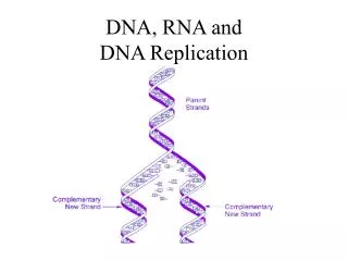
5’ 3’ The two strands of the DNA double helix are COMPLEMENTARY to each other. Means that when oriented with opposite polarity, their nitrogenous bases are in sequence to form perfect Watson-Crick base pairs. Watson and Crick suggested that to replicate DNA, strands were separated by breaking weak hydrogen bonds between base pairs. Each strand then has information to direct synthesis of new complementary strand to form 2 double helices. 5’ 3’ 3’ 5’ 3’ 5’
DNA POLYMERASE (in E. coli, three versions, I, II, and III) Cannot initiate new strand, can only ADD nucleotides to 3’ end of growing strand that are complementary to template strand. Synthesis ALWAYS in 5’ to 3’ direction. Uses triphosphate forms of nucleotides as precursors.