Download
1 / 39
390 likes | 392 Views
Explore the male reproductive system, including the testes, ducts, accessory sex glands, supporting structures, and the process of spermatogenesis. Learn about the hormonal control and functions of the male reproductive system.

E N D
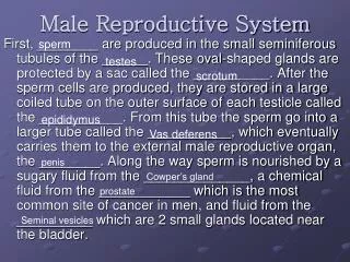
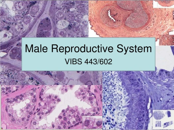
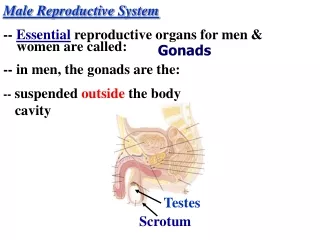
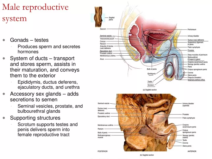
Male reproductive system • Gonads – testes • Produces sperm and secretes hormones • System of ducts – transport and stores sperm, assists in their maturation, and conveys them to the exterior • Epididymis, ductus deferens, ejaculatory ducts, and urethra • Accessory sex glands – adds secretions to semen • Seminal vesicles, prostate, and bulbourethral glands • Supporting structures • Scrotum supports testes and penis delivers sperm into female reproductive tract
Scrotum • Supporting structure for testes • Scrotal septum – internally divides scrotum into two sacs, each with a single testis • Made up of subcutaneous layer and dartos muscle (smooth) • Associated with each testis is the cremaster muscle (skeletal) • Normal sperm production requires a temperature 2-3°C below core body temperature • Cremaster and dartos muscle contracts or relaxes
Testes or testicles • Paired oval glands in the scrotum • Develops near kidney and descends through inguinal canals near 7th month of fetal development • Tunica vaginalis partially covers testes • Tunica albuginea – internal to tunica vaginalis • Extends inward forming septa that divide testis into lobules • Each of 200-300 lobules contains 1-3 seminiferous tubules • Sperm produced here through spermatogenesis
Seminferous tubule cells • Spermatogenic cells – sperm-forming cells • Spermatagonia (stem cell) develop from germ cells that arise in yolk sac and enter testes in 5th week of development spermatocyte spermatidspermatozoa • Sertoli cells– support cells • Tight junction form blood-testis barrier – prevents immune response against sperm cell surface antigens • Nourish spermatocytes, spermatids and sperm, phagocytize excess spermatid cytoplasm, control movements of spermatogenic cells, release sperm into lumen, produce fluid for sperm transport, secrete inhibin, regulate effects of testosterone and follicle-stimulating hormone (FSH) • Leydig (interstitial) cells found in spaces between seminiferous tubules • Secrete testosterone
Spermatogenesis • Takes 65-75 days • Begins with spermatogonia – diploid (2n) • Stem cells undergo mitosis to replace themselves and some continue development • Primary spermatocytes – diploid (2n) • Each duplicates its DNA and meiosis begins • Meiosis I – homologous pairs line up, crossing over occurs • Secondary spermatocytes (haploid or n) • 2 cells at end of Meiosis I • Each chromosome made up of 2 chromatids attached at centromere • Meiosis II – 2 chromatids separate • Spermatids – 4 haploid cells at end of meiosis II • Cells remain attached to each other by cytoplasmic bridges • Spermiogenesis – development of spermatids into sperm • Spherical spermatids transform into elongated sperm • Acrosome and flagella form, mitochondria multiply • Sertoli cells dispose of excess cytoplasm • Spermiation – release from connections to Sertoli cells • Not yet able to swim
Sperm • Each day about 300 million sperm complete spermatogenesis • Head • Nucleus with 23 chromosomes (haploid or n) • Acrosome – vesicle filled with oocyte penetrating enzymes • Tail • Neck – contains centrioles forming microtubules that comprise remainder of tail • Middle piece – contains mitochondria • Principal piece – longest portion of tail • End piece – terminal, tapering portion of tail • Once ejaculated, sperm do not survive more than 48 hours in female reproductive tract
Androgens (testosterone and DHT) • Prenatal development • Testosterone stimulates male pattern of development or reproductive system ducts and descent of testes • DHT stimulates development of external genitalia • Development of male sexual characteristics • At puberty, they bring about development of male sex organs and development of male secondary sexual characteristics • Development of sexual function • Androgens contribute to male sexual behavior, spermatogenesis and sex drive (libido) • Stimulation of anabolism • Stimulate protein synthesis – heavier muscle and bone mass in men
Male reproductive system ducts • Spermatic cord • Ascends out of scrotum • Consists of ductus deferens as it ascends through scrotum, testicular artery, veins that drain testes and carry testosterone, autonomic nerves, lymphatic vessels, and cremaster muscle
Reproductive system ducts in males • Ducts of testis • Pressure generated by fluid produced by Sertoli cells push sperm along seminiferous tubules, straight tubules, rete testis, efferent ducts, then ductus epididymis • Epididymis • Consists of tightly coiled ductus epididymis • Stereocilia are microvilli that reabsorb degenerated sperm • Site of sperm maturation – acquire motility and ability to fertilize • Can store sperm for several months • Continues as ductus (vas) deferens • Ductus (vas) deferens • Conveys sperm during sexual arousal through peristaltic contractions • Can also store sperm several months
Ejaculatory ducts • Formed by union of duct from seminal vesicle and ampulla of ductus deferens • Terminate in prostatic urethra • Eject sperm and seminal vesicle secretions just before release of semen into urethra • Urethra • Shared terminal duct of reproductive and urinary systems • Subdivided into prostatic urethra, membranous urethra, and spongy (penile) urethra • Ends at external urethral orifice
Accessory sex glands – secrete most of liquid portion of semen • Seminal vesicles - About 60% of semen volume • Secrete alkaline, viscous fluid containing fructose, prostaglandins, and clotting proteins (different from blood) • Prostate - About 25% of semen volume • Secretes milky, slightly acidic fluid containing citric acid, several proteolytic enzymes, acid phosphatase, seminalplasmin (antibiotic) • Bulbourethral glands • Secrete alkaline fluid that protects passing sperm by neutralizing acids from urine in urethra • Mucus lubricates end of penis and lining of urethra
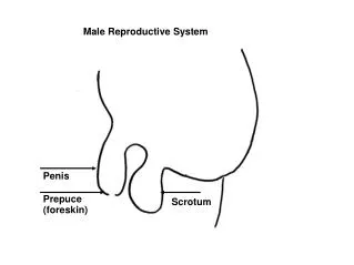
Semen and Penis • Semen • Mixture of sperm and seminal fluid • Typical volume 2.5-5 mL with 50-150 million sperm/mL • Slightly alkaline pH of 7.2-7.7 due to seminal vesicle secretions • Provides transport medium, nutrients, and protection • Coagulates after ejaculation due to clotting proteins • Penis • Contains urethra • Passageway for ejaculation of semen and excretion of urine • Body of penis – 3 cylindrical masses of tissue with erectile tissue • Glans penis – terminal opening is external urethral orifice • Prepuce or foreskin covers glans in uncircumcised men • Root of penis is attached portion • Erection – parasympathetic fibers release and cause local production of nitric oxide (NO) causing smooth muscle in arterioles to relax and dilate allowing large amounts of blood to enter penis
Abnormal conditions of Male reproductive system • Cryptorchidism- “hidden testis” • 3% full term males; 30% premature males • Sterility • Cancer risk30-50x higher • Testicular cancer • Most common cancer between 20-35 • More than 95% arise from spermatogenic cells w/in seminiferous tubules • Dull ache in lower abdomen; testicular heaviness; • Early detection self exams • Gently roll testicle between index finger and thumb • Feel for lumps, swellings, or other changes • Prostate cancer • Leading cause of death from cancer in men in U.S. • Each year 200,000 diagnosed; 40,000 deaths • Prostate Specific Antigen (PSA) is produced only by prostate epithelial cells, increases with enlargement of prostate (many indicate infection, benign growth, or cancer) • Blood test measures level of PSA (recommended for males over 50) • Digital Rectal Exam (palpate gland via rectum)
Vasectomy • Portion of vas deferens is removed • Remaining pieces are stitched closed • Sperm production continues but can not reach exterior • Sperm degenerate & destroyed by phagocytosis • Testosterone levels remain the same • If done correctly 100% effective • Can be reversed; chance of regaining fertility is only 30-40%
Female reproductive system • Gonads – ovaries • Uterine (fallopian) tubes or oviducts • Uterus • Vagina • External organs – vulva • Mammary glands
Ovaries • Paired glands homologous to the testes • Produce • Gametes – secondary oocytes that develop into mature ova (eggs) after fertilization • Hormones including progesterone, estrogens, inhibin and relaxin • Series of ligaments hold ovaries in place • Broad ligament – part of parietal peritoneum • Ovarian ligament – anchors ovaries to uterus • Suspensory ligament – attaches ovaries to pelvic wall
Histology of ovary Ovarian medulla • Contains blood vessels, lymphatic vessels, and nerves • Ovarian cortex • Ovarian follicles - consist of oocytes in various stages of development • Surrounding cells nourish developing oocyte and secrete estrogens as follicle grows • Mature (graafian) follicle – large, fluid-filled follicle ready to expel secondary oocyte during ovulation • Corpus luteum – remnants of mature follicle after ovulation • Produces progesterone, estrogens, relaxin and inhibin until it degenerates into corpus albicans
Oogenesis and follicular development • Formation of gametes in ovary • Oogenesis begins before females are born • Essentially same steps of meiosis as spermatogenesis • During early fetal development, primordial (primitive) germ cells migrate from yolk sac to ovaries • Germ cells then differentiate into oogonia – diploid (2n) stem cells • Before birth, most germ cells degenerate – atresia • A few develop into primary oocytes that enter meiosis I during fetal development • About 200,000 to 2,000,000 at birth, 40,000 remain at puberty, and around 400 will mature • Each month from puberty to menopause, FSH and LH stimulate the development of several primordial follicles • Usually, only one reaches ovulation
Uterine (fallopian) tubes or oviducts • Provide a route for sperm to reach an ovum • Transport secondary oocytes and fertilized ova from ovaries to uterus • Infundibulum ends in finger-like fimbriae • Produce currents to sweep secondary oocyte in • 3 layers • Mucosa – ciliary conveyor belt, cells provide nutrition to ovum • Muscularis – peristaltic contractions • Serosa – outer layer
Uterus • Anatomy • Fundus, body, isthmus, and cervix (opens into vagina) • Normal position is anteflexion – anterior and superior over bladder • Histology – 3 layers • Perimetrium – outer layer • Part of visceral peritoneum • Myometrium • 3 layers of smooth muscle • Contractions in response to oxytocin from posterior pituitary • Endometrium – inner layer • Highly vascularized • Stratum functionalis –lines cavity, sloughs off during menstruation • Stratum basalis – permanent, gives rise to new stratum functionalis after each menstruation
Uterus • Blood supply • Essential to support regrowth after menustration, implantation and development of placenta • Cervical mucus - produced by secretory cells of cervix mucosa (20-60ml/day) • Water, glycoproteins, lipids, enzymes, and inorganic salts • More hospitable to sperm near ovulation – thinner, more alkaline • Supplements energy needs of sperm, protect sperm from phagocytes and hostile environment of tract • Plays a role in capacitation
Fibromuscular canal extending from exterior of body to cervix Mucosa continuous with uterine mucosa Decomposition of glycogen makes acidic environment hostile to microbes and sperm Alkaline components of semen raise pH Muscularis – 2 layers of smooth muscle Adventitia – anchors vagina to adjacent organs Vagina
Vulva – external female genitalia • Mons pubis – cushions pubic symphisis • Labia majora – homologous to scrotum • Contains pubic hair, adipose tissue, sebaceous glands, sudoriferous glands • Labia minora – homologous to spongy (penile) urethra • No hair or fat, few sudoriferous glands, many sebaceous glands • Clitoris – 2 small erectile bodies, corpora cavernosa and numerous nerves and blood vessels • Homologous to glans penis • Vestibule – region between labia minora • External urethral orifice, openings of several ducted glands, and vaginal orifice, Hymen – forms border around and partially closes vaginal orifice • Homologous to membranous urethra of males • Bulb of the vestibule – 2 elongated masses of erectile tissue on either side of vaginal orifice
Perineum • Diamond-shaped area medial to thighs and buttocks of males and females • Contains external genitalia and anus
Breast / Mammary glands • Nipple has openings of lactiferous ducts • Areola – pigmented area; contains sebaceous glands • Mammary gland – modified sudoriferous gland that produces milk • 15-20 lobes divided into lobules composed of alveoli (milk-secreting glands)
Encompasses ovarian and uterine cycle, hormonal changes that regulate them, and related changes in breast and cervix Ovarian cycle – series of events in ovaries that occur during and after maturation of oocyte Uterine (menstrual) cycle – concurrent series of changes in uterine endometrium preparing it for arrival of fertilized ovum The Female Reproductive Cycle
Gonadotropin-releasing hormone (GnRH) Secreted by hypothalamus controls ovarian and uterine cycle Stimulates release of follicle-stimulating hormone (FSH) and luteinizing hormone (LH) from anterior pituitary FSH Initiate follicular growth Stimulate ovarian follicles to secrete estrogens LH Stimulates further development of ovarian follicles Stimulate ovarian follicles to secrete estrogens Stimulates thecal cells of developing follicle to produce androgens to be converted into estrogens Triggers ovulation Promotes formation of corpus luteum – produces estrogens, progesterone, relaxin and inhibin Hormonal regulation
Hormonal regulation • Relaxin • Produced by corpus luteum • Relaxes uterus by inhibiting contraction of myometrium • At end of pregnancy, increases flexibility of pubic symphysis and dilates uterine cervix • Inhibin • Secreted by granulosa cells of growing follicles and by corpus luteum • Inhibits secretion of FSH and LH
Secretion and physiological effects of hormones in the female reproductive cycle