Download
1 / 33
420 likes | 760 Views
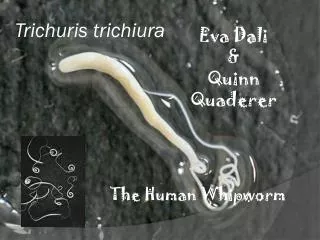
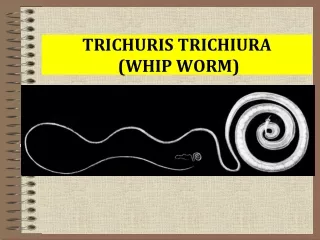
Trichuris trichiura Whip worm. Common name: whip worm Disease: trichuriasis, whip worm infection Final host: human, dogs, pig, monkey Habitat: large intestinal ( caecum, appendix, rectum) Geographical distribution: Cosmopolitan with poor sanitation.

E N D
Common name: whip worm • Disease: trichuriasis, whip worm infection • Final host: human, dogs, pig, monkey • Habitat: large intestinal ( caecum, appendix, rectum) • Geographical distribution: Cosmopolitan with poor sanitation. • Children are more likely to be infected than adults because they are more likely to have close physical contact with contaminated soil
Infective stage: infective larva in egg • Transmission occurs through ingestion of eggs, usually on contaminated vegetables or soil. • Diagnostic stage: Egg barrel shape with polar plugs
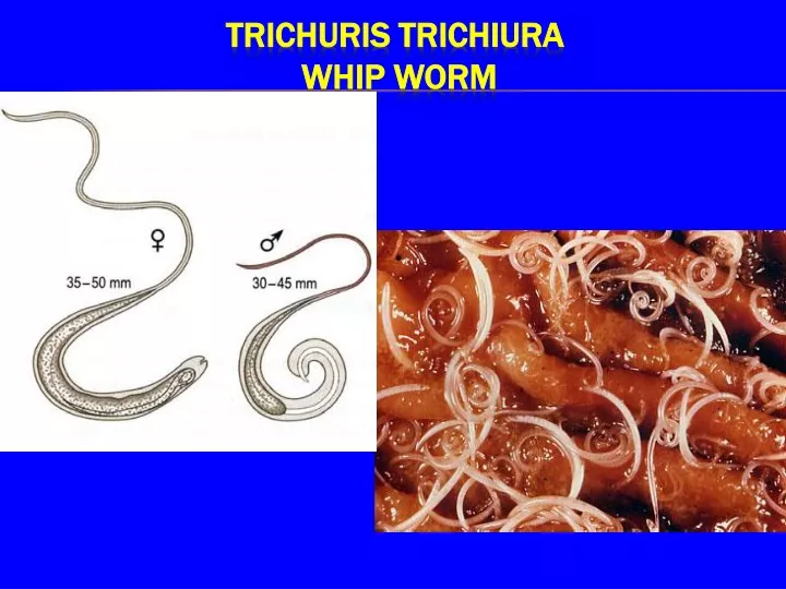
Adult female worm The anterior two-thirds of the body being very thin (looks like a whip) and the remaining posterior end is thick and linear. Size: 3.5-5cm in length Adult Male worm smaller than the female, 3.0-3.5cm. The posterior end is curved and has a single spicule enveloped with sheath. Morphology:
The anterior end two-thirds of the body being very thin (looks like a whip). • Adult worm penetrates into and embed its whip-like anterior portion in the intestinal mucosa, By small spear Adult male Adult female • Longer than the male.- posterior end is thick • and linear. • Shorter than the female. posterior end curved and • has a single spicule • enveloped with sheath. • .
posterior end curved and has a single spicule enveloped with sheath
Eggs: Shape: barrel–shaped Size: 50-55 x 25-30μm Shell: thick egg shell with 2 polar plugs Color: Yellow-brown Content: immature egg cells 3000-10000 eggs daily output
Eggs pass out immature Embryo develops inside the egg (that takes about 3weeks at 25C) Mature eggs swallowed 1st stage larvae hatch in small intestine and penetrate villi Then migrate to large intestine and attach to mucosa with the thin anterior end After 2-4 month females mature and lay eggs. Life cycle
Pathology: • Light infection with Trichiuris are asymptomatic • Heavier infections are characterized by 1- diarrhea, 2- anorexia, 3- nausea 4- abdominal pain 5- anemia may be the result of hemorrhaging when the worms mucosal damage))penetrate the intestinal wall • Rectal prolapse. Children’s infection can cause rectal prolapse, The reason is the cecum is damaged by the worm, the cecum can be pushed out from the anus.
Pathology of Trichuris trichiura (worms cause loss of muscle tone in wall of rectum and it everts out the anus; whipworms are often seen attached to the rectal tissue
1- Eggs or worm in feces. Eggs are oval, barrel shaped, 2- Eosinophilia may occur. 3- In heavy infection proctoscopy or sigmoidoscopy, can show the worms attached to the mucosa. 4- Visual detection of adult worms on prolapsed rectum. Laboratory diagnosis
Common name: Trichina worm- The Pork worm • Trichinella spiralis means spira, how this coils up in its host. • diseases: Trichinosis, Trichiniasis, • is a zoonotic disease. it is passed between humans and animals. • Habitat: females in mucosa of small intestinal • The same animal acts as final and intermediate host • is most common in Europe, North America, and Asia, • Infective stage: contaminated meat (muscle) containing encyst larvae (pig) • Diagnostic stage: larvae encysted in muscle (human) • Can be fatal if large numbers of cysts form in the heart muscle. Trichinellaspiralis
Morphology: Anterior end: slender with small non papilated mouth. Male:1.5mm posterior end ventrally curved with 2 copulatory appendages Female: 3.5mm single ovary Posterior end bluntly rounded
The sperm fertilizes the egg, but the female doesn’t release the eggs until they have hatched within her uterus, which is usually within the fifth or sixth day of infection. Female worms can produce approximately 1500 newborn larvae (immature L1) during a life span (4-16 weeks), before expulsion by the host immune system. Viviparous: Expel active larvae
Oviparous: Eggs laid in 1-cell stage or early cleavage stage. Ovoviviparous: Eggs laid containing embryo or larva (L1). Some hatch out prior to passage in feces. Viviparous: Expel active larvae
LARVAE Measures 80-120 um by 5.6 u at birth And grows but little until it has entered A muscle fiber, size 900 um developed.
Female worms deposit larvae into: The mucosa of small intestine Directly into the lymphatics Blood stream. The larvae carried to all parts of body, but the larvae developing only in striated muscle.
Epidemiology • The disease among humans, rats, and pigs • Rats and pigs feeding on garbage that includes infected pork waste, become infected in turn • Dead or dying infected rats are themselves eaten by pigs • Raw or poorly cooked pork (sausage) harboring infective larvae then becomes the vehicle for human infections • Trichinellosis is a cosmopolitan disease that occurs most commonly in Europe and the US
Pathogenesis: 1--Penetration of the adult females into mucosa The first symptoms appear between 1- 2 days after ingestion. The worms migrating in the intestinal epithelium Inflammation of duodenal and jejunal mucosa: This causes: inflammation, nausea, vomiting, sweating, and diarrhea.
2- The migrating larvae Ten days after infection the larvae will penetrated the muscle fibers, carried to all parts of body, This causes • muscular pain, • difficulty breathing • Per orbital edema and conjunctivitis • heart (myocarditis), • lungs (pneumonitis), • brain (encephalitis). • Can be fatal if large numbers of cysts form in the heart muscle. heart failure or respiratory or kidney malfunction
Diagnosis : • Muscle biopsy at the encystment stage • blood test for eosinophilia • increased levels of creatine phosphokinase CPK, • Serology test Immunoassays, such as ELISA • At the diarrheal stage, adult and larvae may be found in feces
Treatment • Anti-inflammatory drugs. When infected, it is also suggested that you get a lot of rest to help give your body time to recover. • Mebendazole is the drug of choice.