Download
1 / 49
520 likes | 649 Views
Medical Imaging. X-Rays I. Principle of X-ray. A source of radiation. Principle of X-ray. A source of radiation. A patient of non uniform substance. Principle of X-ray. A source of radiation. A shadow. A patient of non uniform substance. Principle of X-ray. A source of radiation.

E N D
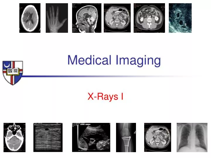
Medical Imaging X-Rays I
Principle of X-ray A source of radiation
Principle of X-ray A source of radiation A patient of non uniform substance
Principle of X-ray A source of radiation A shadow A patient of non uniform substance
Principle of X-ray A source of radiation
X-ray tube • Working Principle: Accelerated charge causes EM radiation: • Cathode filament C is electrically heated (VC = ~10V / If = ~4 A) to boil off electrons • Electrons are accelerated toward the anode target (A) by applied high-voltage (Vtube = 40 – 150 kV); • Deceleration of electrons on target creates "Bremsstrahlung" evacuated gas envelope filament VC, If + A - C - +
X-ray tube • Cathode Filament (-) • Coil of tungsten wire • High resistance in coil ->temperature rise to > 2200oC • Thermionic emission of electrons • Tube (vacuum) • Typical: Vtube = 40 – 150 kVp, Itube = 1-1000mA evacuated gas envelope filament VC, If - + - - - - - A - - - - C space charge stops further emission kVp, Itube - +
X-ray tube • Anode • Tungsten (high atomic number Z=74) • Electrons striking the anode generate HEAT and X-Rays • In mammography ->Molybdenium (Z=42) and Rhodium (Z=45) • Stationary anode-> tungsten embedded in copper • Rotating anode (3000 to 10,000rpm) -> increase heat capacity, target area evacuated gas envelope filament VC, If - + - - - - - A - - - - C space charge kVp, Itube - +
X-RAY production • X-ray tube produces two forms of radiation • Bremsstrahlung radiation (white radiation) • Characteristic radiation
White radiation, Bremsstrahlung (Brake) • Inelastic interaction with atoms nuclei • Loss of kinetic energy • Xray (E) = lost kinetic E X-Ray • High kinetic energy • Forward radiation • Emission Z2 electron Coulombic interaction (Atomic number) # of protons
White radiation, Bremsstrahlung -Smaller L produce larger X-ray -Broad range of emitted wavelengths X-Ray L
How many wavelength will be emitted by a beam of electrons underegoing “Bremsstrahlung ”
White radiation, Bremsstrahlung -Smaller L produce larger X-ray -Broad range of emitted wavelengths X-Ray L impact with nucleus maximum energy
X-ray intensity -QUANTITY • Overall Bremsstrahlung intensity I : • 90% of electrical energy supplied goes to heat, 10% to X-ray production • X-ray production increases with increasing voltage V
Bremsstrahlung spectrum relative output • Theoretically, bremsstrahlung from a thick target creates a continuous spectrum from E = 0 to Emax • Actual spectrum deviates from ideal form due to • Absorption in window / gas envelope material and absorption in anode • Multienergetic electron beam Peak voltage kVp
Characteristic radiation relative output • Energy must be > binding energy • Discrete energy peaks due to electrons transitions • Ka transition L->K • Kb transition M,N,O->K Peak voltage kVp
Characteristic radiation Incident electron
Characteristic radiation l2 Incident electron Occurs only at discrete levels There is a possibility of forming Auger electrons
Characteristic radiation • In Tungsten characteristic X-ray are formed only if V>69.5 kV because K shell binding energy is 69.5 keV • Molybdenum K-shell can be obtained at V> 20kV • L shell radiation is also produced but it’s low energy and often • absorbed by glass enclosure
X-ray intensity -QUALITY • Effective photon energy produced • Effective = ability to penetrate the patient • Effective photon energy ~ 1/3 to ½ of energy produced • Higher energy better penetration • Beam filtration – beam hardening
Beam Hardening Polyenergetic beam ------------------------------->monoenergetic beam
Anode Most of the energy deposited on the anode transfers into heat
Reduction of anode heating • Made of Tungsten, high melting point high atomic number Z = 74 Kinetic energy of incident electrons 100keV electron 6 MeV electron
Anode • the target angle, 7 to 20 (average 12) Seffective = Sactual*sin() -----------> Line focusing principle
Anode filament balance General radiography
Heel effect - SID source to image distance - Heel effect is smaller at smaller SID Reduction of intensity on the anode side SID The reduction in intensity can be used to reduce patient exposure
Beam collimation • Size and shape of the beam • Lead shutters • Dose reduction
Reduction of anode heating • Anode angle of 7º…15º results in apparent or effective spot size Seffectivemuch smaller than the actual focal spot of the electron beam (by factor ~10) • Rotation speed ~ 3000 rpm • Decreases surface area for heat dissipation from by a factor of 18-35.
Limitations of anode angle • Restricting target coverage for given source-to-image distance (SID) • "Heel effect" causes inhomogeneous x-ray exposure
X-ray tube - space charge • Space charge cloud forms at low tube voltage • At low filament current a saturation voltage is achieved, rising tube voltage will not generate higher electron flow • At high filament current and low tube voltage, space charge limits tube current->space-charge limit
Space charge limited • At high filament current and low tube voltage, space charge limits tube current->space-charge limit
Generator • Single phase • Single phase input (220V, 50A) • Single pulse or double pulse->rectifier • Min exposure time 1/120 sec • Xray tube current non linear below 40kV • Three phase • Three phase wave, out of phase 120 deg • More efficient higher voltage • Better control on exposure
Rectifier Protects cathode from anode thermionic emission
Rectifier 1 phase 3 phase
Principle of X-ray A source of radiation A patient of non uniform substance
N = Noe-mL Attenuation N True for monoenergetic x-ray Loss of photons by scattering or absorption L1 L No N m -> linear attenuation coefficient L1
m linear attenuation coeff. • m = mr+ mph+ mc+ mp [cm-1] • rayleigh • photoelectric • Compton • pair
m linear attenuation coeff. • m = mr+ mph+ mc+ mp [cm-1] • depends on tissue • soft tissue, hard tissue, metals • mdecreases when energy increase • soft tissue: • m = 0.35 0.16 cm-1 for E = 30 100keV • mdepends on density of material • mwat > mice> mvapor
Mass attenuation coeff. N = Noe- r(m/r)L rL = mass thickness
I x Mass attenuation coeff. N = Noe- r(m/r)L rL = mass thickness
Poly-energetic beam • Mass attenuation coefficient and linear attenuation coefficient are for mono-energetic beam • Half-value layer is for quantifying poly-energetic beams
HVL half value layer • Thickness of material attenuating the beam of 50% - narrow beam geometry • HVL for soft tissue is 2.5 3.0 cm • at diagnostic energies
HVL half value layer • Transmission of primary beam: • 10% chest radiography • 1% scull radiography • 0.5% abdomen radiography • Mammography (low energy HVL 1 cm)
Mean free path 1/m • Average distance traveled before interaction MFP=1/m HVL mfp
Principle of X-ray A source of radiation A shadow A patient of non uniform substance