Download
1 / 29
400 likes | 3k Views
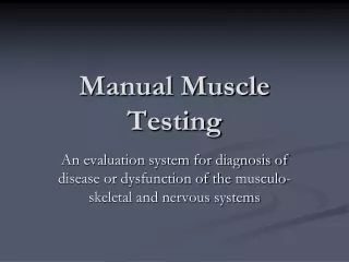
Manual Muscle Testing. An evaluation system for diagnosis of disease or dysfunction of the musculo -skeletal and nervous systems. Purpose. Measures the capability of muscles or groups to provide support and movement Diagnostic tool Postural balance Gait impairment Range of motion

E N D
Manual Muscle Testing An evaluation system for diagnosis of disease or dysfunction of the musculo-skeletal and nervous systems
Purpose • Measures the capability of muscles or groups to provide support and movement • Diagnostic tool • Postural balance • Gait impairment • Range of motion • Uses little equipment • Obtains information not defined by other procedures
Precautions • Do No Harm (use gentleness) • Know ROM limits • Follow procedure • Record • Promptly • Accurately
To Get Standardized Results • Proper training and education • Knowledge base of anatomy, physiology and neurology of muscle function • Follow precise testing protocol • Practice, Practice, Practice • A skill developed and maintained with number of cases
Validity and Accuracy Coordinate the muscle testing findings with other standard diagnostic procedures The amount of pressure used to test may vary between persons performing the test. The amount of strength loss must be greater than approximately 20to 30% to be dependably measurable Comparison of both sides is a better indicator of loss
Muscles • 3 Types: Skeletal, Smooth, Cardiac • Skeletal around 40% of muscle composition • Generally voluntarily controlled • Composed of fibers • Work in groups • Movement depends on how the muscles are attached
Structure of Muscle http://en.wikibooks.org/wiki/Anatomy_and_Physiology_of_Animals/Muscles
How Do Muscles Cause Movement Origin- where the muscle is attached to the bone; this bone will move very little Insertion- muscle attachment to bone with most motion Belly of muscle- part of muscle that enlarges on contraction
Muscle Groups Trapezius Latisimusdorsi Deltoids Biceps Triceps • Quadriceps • Hamstrings • Calf • Low back • Abdominals • Pectoralis major • Rhomboids
Conduct Strength Testing • Correct positioning is essential (Start in extended anatomical position) • Place muscle to be tested in a supported position directly opposed to gravity • Exert uniform force directly on the line opposing movement
Testing of Bicep & Tricep • Support humerus where gravity is against the bicep and tricep, client in anatomical position • Move elbow through full ROM (Passive ROM) • Flexion • Extension • Internal rotation • External rotation
Maneuver to Assess Muscle Strength With arm in full extension, pull downward on forearm while client attempts to flex. With arm flexed, apply pressure against forearm, ask client to straighten arm. When performing muscle tests, be sure to evaluate for asymmetry of the muscle groups (i.e. atrophy on one side and not the other) and landmarks prior to testing.
Use the following scale to rate strength: 0-No movement, no contraction of the muscle 1- Trace, evidence of muscle contraction but no joint movement 2- Poor, complete range of motion with gravity eliminated 3-Fair, complete range of motion against gravity 4- Good, complete range of motion against gravity with moderate resistance 5-Normal, complete range of motion against gravity with maximal resistance without evidence of fatigue
Other Test Results • Weakness – defined as a strength below fair in non weight bearing muscles; below fair + in weight bearing muscles • Contracture – degree of shortness in muscle, so it cannot move through ROM • Substitution – weak muscles are supported by other muscles to move
Active ROM • Instruct client to move the elbow through ROM • Flexion • Extension • Internal rotation • External rotation • Normal ROM is measured by goniometer • Elbow flexion 0-160 • Elbow extension 145-0 • Elbow pronation (rotation inward) 0-90 • Elbow supination (rotation outward) 0-90
Strength Test Example • Gastrocnemius (Ankle plantar flexion) • Patient Standing • Rises on toes, pushing weight upward
Case Study to Follow Take patient hx to determine diagnosis Assessment of muscle strength Set objectives Implement a plan Evaluate progress
Case Study • Drop Foot • weakness of muscles that are involved in flexing the ankle and toes.
Clinical Muscle Evaluation • Typical podiatric ankle strength evaluation consists of plantar flexion, dorsiflexion, eversion and inversion testing
Dorsiflection Testing • Tibialis Anterior • Support leg above ankle • Apply pressure against medial side, dorsal surface of the foot, in the direction of plantar flexion of the ankle joint and eversion of the foot. Test dorsiflection directly.
Dorsiflection Testing • Extensor Hallucis Longus • Stabilize foot in slight plantar flexion • Pressure applied against dorsal surface of distal and proximal phalanges of the great toe in direction of flexion. Test big toe extension.
Dorsiflection Testing • Extensor Digitorum Longus • Stabilize foot in slight plantar flexion • Apply pressure against dorsal surface of the toes in the direction of flexion. Test extension of toes.
Summary • Manual Muscle Testing is clinical tool used to evaluate patient • Need information in order to develop orthotic treatment plan
END • Questions?
Conditions A list of conditions treatable with Applied Kinesiology
All about muscles http://www.emporia.edu/ksn/v42n1-january1996/shape.htm
This power point is based on information found on the Illinois Institute of Technology web site where students developed resources to be used in education in Latin America. I modified the original power point to be used by high school students in the Healthcare Science classroom. Pat Rape
http://www.lhup.edu/yingram/jennifer/webpage/homepage2.htm http://www.iit.edu/~ipro309s08/links.html http://www.bulowbiotech.com/intromov.html
Careers http://francistuttle.com/classes/ctp/details.aspx?PRGID=13 Orthotic & Prosthetic (O & P) Technicians assist the disabled by fabricating the orthopedic braces (orthoses) and artificial limbs (prostheses) necessary for their rehabilitation. O & P Technicians are trained and skilled to provide comprehensive O & P technical support services and possess the knowledge to interact with clinical prosthetists and orthotists. You will acquire knowledge in polymer processes, strength of materials and applied biomechanical principles to develop and totally customize an orthosis or prosthesis. Providing O & P care involves the application of clinical and technical processes to meet patient rehabilitation objectives.