Download
1 / 47
490 likes | 646 Views
Unit 4: Nervous System Lab 3: Cranial/Spinal Nerves & Lab 4, Part 1: Introduction to the Eye. Jessica Radke -Snead, RD, MS Bio 241 Anatomy & Physiology. Reminders. Today’s Lab Cranial and spinal nerves Introduction to Lab 4 Wednesday Human eye/vision Histology of the retina

E N D
Unit 4: Nervous SystemLab 3: Cranial/Spinal Nerves & Lab 4, Part 1: Introduction to the Eye Jessica Radke-Snead, RD, MS Bio 241 Anatomy & Physiology
Reminders • Today’s Lab • Cranial and spinal nerves • Introduction to Lab 4 • Wednesday • Human eye/vision • Histology of the retina • Human ear and physiological aspects of hearing • Histology of the cochlea • Visual acuity tests (observe on your own) • Hearing tests (observe on your own)
I Olfactory Nerve Type: Sensory Function: Smell (olfaction) Olfactory receptors (olfactory epithelium) extend through olfactory foramina (cribiform plate, ethmoid bone) and collectively form the right and left olfactory nerves and olfactory bulbs. Axons of the olfactory tracts end in the primary olfactory area (temporal lobe).
II Optic Nerve Type: Sensory Function: Vision • Eye, rods and cones initiate visual signals • Signals relayed via optic nerve, which merge to form the optic chiasm (optic foramina) and optic tracts (posterior) • Axons from optic tracts project to the primary visual area (occipital lobe)
III Oculomotor, IV Trochlear and VI Abducens Nerves Type: Motor axons that exit the brain stem. Sensory axons (extrinsic eyeball muscles) initially travel through each of these nerves and enter the midbrain (V Trigeminal nerve). Function: Pass through Sup Orb Fissure to control the muscles that move the eyeballs. Sensory: proprioception (non-visual perception)
III Oculomotor“eye-mover” • Type: Motor and Sensory • Extends from midbrain (junction with pons) • Somatic motor axons innervate: • Extrinsic eyeball muscles (SR, MR, IR & IOb) Eyeball movement • Levatorpalebraesuperioris raising the upper lid • Autonomic motor axons innervate intrinsic eyeball muscles (Ciliary) adjust the shape of the lens and iris to adjust the size of the pupil
IV Trochlear Nerve“pulley” • Type: Motor and Sensory • Posterior side of midbrain, wraps around the pons • Somatic motor axons innervate extrinsic muscle (SOb) movement of the eyeball • Trochlea: cartilage that supports the “pulley” action of the SOb, thus nerve is called “trochlear”
VI Abducens Nerves“Away to lead” • Type: Motor and Sensory • Originates in the pons • Somatic motor axons innervate extrinsic muscle (LR) lateral rotation of eyeball • “ABDuction” or lateral movement of eye = ABDucens nerve
V Trigeminal Nerve“triple branches” Type: Motor and Sensory Function: Sensory axons • Touch, pain and temp pons • Proprioceptors in the jaw Motor axons supply jaw muscles for mastication 3 Branches: • Ophthalmic (exits through Sup Orb Fissure) • Maxillary (exits through foramen rotundum) • Mandibular (exits through foramen ovale)
V Trigeminal Nerve“triple branches” • Ophthalmic nerve • Sensory axons from the upper eyelid, eyeball, lacrimal glands, nasal cavity, nose, forehead and anterior scalp • Maxillary nerve • Sensory axons from the nose, palate, upper mouth and lower eyelid • Mandibular nerve • Sensory axons from the anterior tongue (not taste), cheek, skin over the jaw and side of the head and lower mouth
VII Facial Nerve Type: Motor and Sensory Sensory axons: • Touch, pain and temperature from ear canal • Proprioception from face and scalp muscles Motor neurons (pons) innervate facial, scalp and neck muscles for facial expression Autonomic motor neurons innervate lacrimal and salivary glands
VIII Vestibulocochlear Nerve“small, spiral/snail like” Type: Sensory • 2 Branches • Vestibular: impulses for equilibrium from the semicircular canals, saccule and utricle of the inner ear to the pons and cerebellum • Cochlear: impulses for hearing from the spiral organ of the inner ear to the primary auditory area (temporal lobe)
IX Glossopharyngeal Nerve“tongue, throat” Type: Motor and Sensory Sensory axons • Arise from taste buds (posterior tongue) and external ear to convey touch, pain and temperature • Proprioceptors in swallowing muscles • Neck region: info from baroreceptors (carotid sinus) that monitor BP and chemoreceptors (carotid bodies) that monitor blood gas Motor axons • Arise in medulla oblongata, pass through the jugular foramen to innervate muscle in pharynx for swallowing • Autonomic axons stimulate the parotid gland (saliva)
X Vagus Nerve“vagrant, wanderer” Type: Motor and Sensory Sensory axons arise from: • External ear: touch, pain and temperature • Taste buds (throat) • Proprioceptors in muscles of the neck and throat • Carotid sinus: monitor BP • Carotid body and aortic bodies: monitor blood gas levels • Organs (thoracic, abdominal cavities): hunger, fullness and discomfort Sensory axons pass through jugular foramen Medulla oblongata
X Vagus Nerve“vagrant, wanderer” Type: Motor and Sensory Motor neurons: muscles of the pharynx and larynx for speech and swallowing Autonomic motor neurons: lungs, heart and smooth muscle and glands of the respiratory passageways and GI tract
XI Accessory Nerve“assisting” Type: Motor and Sensory Motor axons • Arise in the anterior gray horn of the spinal cord (cervical portion) • Exit the spinal cord laterally • Enter the foramen magnum • Exit through the jugular foramen (along with IX and X) why it’s considered a cranial vs spinal nerve Convey impulses to the SCL and trapezius muscles coordinate head movements
XI Accessory Nerve“assisting” Type: Motor and Sensory Sensory axons arise from • Proprioceptors in the SCL and trapezius muscles • Eventually join nerves at the cervical plexus Enter the spinal cord Medulla oblongata
XII Hypoglossal Nerve Type: Motor and Sensory Motor neurons originate in the medulla oblongata hypoglossal canal tongue muscles for speech and swallowing Sensory axons originate from proprioceptors in the tongue muscles extend toward the brain (hypoglossal nerve) then leave hypoglossal nerve to join the cervical spinal nerves medulla oblongata
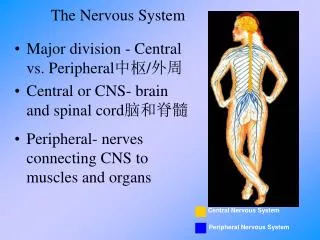
Spinal Cord and Spinal Nerves • Posterior root + Anterior root = spinal nerve at the vertebral foramen • Posterior root contains sensory axons • Anterior root contains motor axons
Spinal Nerves • Spinal cord ends near the L2 vertebrae • Roots of the lumbar, sacral and coccygeal nerves descend at an angle vertebral foramina (caudal equina) • 31 pairs of spinal nerves • Identified by the region and level of vertebral column from which they emerge • 8 pairs cervical • 12 pairs thoracic • 5 pairs lumbar • 5 pairs sacral • 1 pair coccygeal
Anatomy of Spinal Nerves • Upon passing through its intervertebral foramen, a spinal nerve divides into branches (rami) • Posterior ramus: serves the muscles and skin of the posterior trunk • Anterior ramus • Muscles and structures of the upper and lower limbs • Skin of the lateral and anterior trunk • Meningeal branch: vertebrae, spinal cord and meninges • Rami communicantes: components of the ANS Rami communicantes
Plexuses • Network of axons formed on both sides of the body by joining with various numbers of axons • Names often describe the general regions they serve or the course they take • Primary plexuses • Cervical • Brachial • Lumbar • Sacral
Thoracic nerves T2-T12 follow each rib laterally and do not form plexuses
Cervical Plexus • Formed via C1-C4, part of C5, nerves • Supplies: • Skin and muscles on the head, and neck • Superior part of the shoulders and chest • Important nerve arising from this plexsus • Phrenic (spinal nerves C3-C5) • Motor nerve: Diaphragm
Brachial Plexus • Formed by C5-C8 and T1 spinal nerves • Provides majority of the nerve supply to the shoulders and limbs • Complex structure: Roots Trunks Divisions Cords Branches
Brachial Plexus • From the cords, important nerves are: • Axillary (C5-C6) • Motor nerve: Deltoid and Teresminor • Sensory nerve: Lateral arm to the deltoid tuberosity • Radial (C5-T1) • Motor nerve: Triceps, Supinator, Brahcioradialis • Sensory nerve: Posterior arm and forearm, medial side of posterior hand • Median (C5-T1) • Motor nerve: Pronator teres and Flexor carpi radialis • Sensory nerve: Palmar aspect of 2nd-4th fingers • Ulnar (C8-T1) • Motor nerve: Flexor carpi ulnaris • Sensory nerve: Medial portion of 4th and entire 5th finger
Lumbar Plexus • Formed by L1-L4 spinal nerves • Supplies the • Anterior and lateral abdominal wall • External genitals • Part of lower limbs
Lumbar Plexus • Important nerves arising from this plexus are: • Femoral (L2-L4) • Motor nerve: Iliacus, Pectineus, Quadriceps femoris and Sartorius • Sensory nerve: Skin of the lateral anterior thigh and dorsum of the foot • Genitofemoral (L1-L2) • Motor nerve: Cremaster muscle • Sensory nerve: Skin of the medial and anterior thigh, scrotum (male) or labia major (female)
Sacral Plexus • Formed by L4-S4 spinal nerves • Supplies the buttocks, perineum and lower limbs • Important nerves that arise are • Pudendal • Motor nerve: Ischiocavernosus, Bulbospongiosus, Levatorani and External anal sphincter • Sensory nerve: Skin of the penis and scrotum, clitoris, labia major and minora, vagina • Sciatic (posterior) • Branches into tibial and fibular nerves • Motor nerve: Semimembranosus, Semitendinosus, Biceps femoris, Adductor magnus • Sensory: Lateral posterior leg, lateral aspect and plantar surface of the foot
Introduction to the Human Eye • Surface anatomy • Extrinsic and intrinsic eye muscles • Layers forming the posterior wall of the eye • Internal anatomy of the eye • Posterior/vitreous cavity • Anterior cavity
Review on your own • Extrinsic eye muscles • Medial rectus, Superior rectus, Lateral rectus, Inferior rectus, Superior oblique, Inferior oblique and Levatorpalpebraesuperioris • Intrinsic eye muscles • Via Oculomotor nerve (cranial) • Circular iris muscle: (parasympathetic; bright light) pupil constriction • Radial iris muscle: (sympathetic; dim light, action) pupil dilation • Ciliary muscle: smooth muscle that changes the tightness of zonular fibers to change shape of the lens
Surface anatomy of the Eye Tarsal plate
Surface Anatomy of the Eye: Eyelids • Eyelid/Palpebrae • Tarsal plate: elongated dense CT that forms the shape of each palpebrae • Levatorpalpebraesuperioris: raises the upper lid • Oribicularis oculi: closes the eyelid • Superior and inferior palpebral sulci: upper and lower lid creases • Conjunctiva: protective mucous membrane • Palpebral: lines each eyelid • Bulbar: from eyelids into the anterior surface of the eyeball and covers the sclera
Structures of the Eyeball • Fibrous tunic (avascular outer layer) • Cornea: admits and refracts light • Sclera: provides shape and protection • Vascular tunic (middle layer) • Iris: regulates amount of light that enters the eyeball • Ciliary body: secretes aqueous humor and alters shape of lens for near/far vision • Choroid: provides blood supply and absorbs scattered light (melanin)
Structures of the Eyeball • Retina (inner layer) • Receives light receptor potentials and nerve impulses • Provides output to the brain via axons (ganglion cells) optic nerve • Macula lutea: highly pigmented spot near the center of the retina • Fovea centralis: small pit that contains cones (color vision); area of highest visual acuity (sharpness) • Move head and eyes to place images on this point • No rods—periphery of retina; more light sensitive—why you can see a dim star better if you look just to the side of it
Internal anatomy of the Eye • Lens: refracts light and focuses images on the retina to facilitate vision • Posterior/vitreous cavity (lens to wall of retina) • Vitreous body helps maintain the shape of the eyeball and supports retinal attachment to the eyeball • Anterior cavity (cornea to lens) • Aqueous humor helps maintain shape of eyeball and supplies oxygen and nutrients to lens and cornea
Objectives • Today’s Lab 3 • Cranial and spinal nerves • Surface anatomy of the eye • Intrinsic muscles of the eye • 3 layers of the eye • Internal anatomy of the eye • Lacrimal apparatus (if time permits) • Wednesday • Human eye/vision • Histology of the retina • Human ear and physiological aspects of hearing • Histology of the cochlea • Visual acuity tests (observe on your own) • Hearing tests (observe on your own)