Download
1 / 42
420 likes | 704 Views
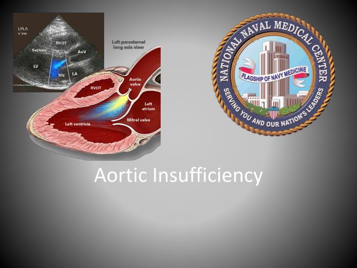
Aortic Insufficiency. Outline. Epidemiology Etiology Pathophysiology Clinical Presentation / Symptoms Natural History Diagnosis Physical Exam Echocardiography Treatment Medical Surgical What’s new?. Epidemiology. Prevalence Framingham Offspring Study 13% in men; 8.5% in women

E N D
Outline • Epidemiology • Etiology • Pathophysiology • Clinical Presentation / Symptoms • Natural History • Diagnosis • Physical Exam • Echocardiography • Treatment • Medical • Surgical • What’s new?
Epidemiology • Prevalence • Framingham Offspring Study • 13% in men; 8.5% in women • Advanced age and male gender associated with AR • Singh,et al. American Journal of Cardiology.1999:83:897-902 • Strong Heart Study (Native American Population) • 10% prevalence • Advanced age and aortic root diameter associated with AR • Lebowitz, et al. JACC. 2000;36:461-7. • In both studies the majority of cases were mild
Etiology • Valvular vs. Aortic Root • Valvular • Calcific AS in the elderly • Infective endocarditis • Congenital bicuspid • More commonly stenosis • Incomplete closure/prolapse can lead to isolated regurgitation • Rheumatic fever • Cusps become infiltrated with fibrous tissue and retract preventing cusp apposition during diastole • Less Common • Congenital: quadricuspid • Inflammatory conditions: SLE, RA, AS, Whipple’s Disease, Takayasu • Anorectic drugs
Etiology • Aortic Root Disease • Age related (degenerative) aortic dilation • HTN longstanding • HLD • Cystic medial necrosis (+/- Marfans) • Aortic dilation related to bicuspid valve • Osteogenesis imperfecta • Syphilitic aortitis • AS, Bechets, Psoriatic arthritis, GCA
Pathophysiology • Regurgitant flow produces increase in LVEDV • Thereby raising wall tension (via Lapace’s law) • Wall stress proportional to (Intraventricular pressure x radius)/wall thickness • LV responds by compensatory eccentric hypertrophy of myocytes • Replication in series and elongation of myocytes and myocardial fibers (Different from AS) Dilation • Chronic compensated AR: sufficient wall thickening occurs so that ratio of wall thickness to cavity radius remains normal. • Maintains End diastolic wall stress normal
Pathophysiology (con’t) • During chronic compensated AR LV is able to adapt to increase in diastolic volume without increasing LV EDP. • LV produces a larger total SV to compensate for regurgitant flow • Over time progressive interstitial fibrosis reduces LV compliance leading to chronic decompensated phase. • Chronic volume overload results in impaired LV emptying, increasing LVEDP and LVEDV causing further dilation and decreasing EF
Pathophysiology • Patients with severe chronic AR have the largest LVEDV of those with any form of heart disease (cor bovinum) • In contrast to AR, AS has pressure overload induced hypertrophy (concentric) with replication in parallel • This leads to increased ratio of wall thickness to radius
Symptoms • Exertionaldyspnea • Orthopnea • Parosxysmal nocturnal dyspnea • Angina pectoris • Late in clinical course • Uncomfortable awareness of heartbeat • Laying down • Thoracic pain • PVCs great heave of volume loaded LV during post extrasystolic beat cause distress • Complaints may be present for many years before symptoms of overt LV dysfunction manifest
Natural History of Chronic AR • Asymptomatic patients with normal EF • No large scale studies evaluating this population • Current ACC recs derived from 9 published series involving 593 patients with mean F/U 6.6 years • Progression to symptoms and/or LV dysfunction: <6%/yr • Progression to asymptomatic dysfunction: <3.5%/yr • Sudden Death: <0.2%/yr • Two multivariate analysis identified two independent predictors of outcome (symptoms, death of LV dysfunction.) • Age • LVEDV • EDV > 50 mm 19% annual • EDV 40-49 mm 6%/yr • EDV < 40 mm <1%/yr
Natural History of Chronic AR • Patients with asymptomatic LV Dysfunction • Most develop symptoms requiring AVR in 2-3 years • Symptom onset 25% annually • Patients with symptomatic AR • No long term studies on this population as most proceed to AVR • Mortality rate of 10% annually if pt has angina • Mortality rate of 20% annually if pt has heart failure Bonow, Circulation 1991, 84:1296-302
Physical Exam • Palpation • Apical impulse enlarged, displaced lateral to midclavicular line in 5th intercostal space • Diastolic thrill and systolic thrill in second intercostal space (increased aortic flow) • Auscultation • Diminished S1 (prolonged PR, LV dysfunction, preclosure of MV) • S2 soft, maybe paradoxically split • S3 may be heard with LV dysfunction (indicating increased LV EDP) • S4 often present (LA contraction into poorly compliant LV) • Blowing diastolic decrescendo murmur starting immediately after A2. • LUSB, diaphragm of stethoscope • Sitting up and leaning forward • Full expiration • Severity correlates with length/duration not intensity of murmur • May hear second diastolic murmur at apex in severe AR • Austin Flint: middle to late diastolic rumble • May hear short midsystolic ejection murmur at base radiating to neck reflecting increased ejection rate (don’t confuse with AS) • Maneuvers • Increase: isometric exercise, squatting, inotrope infusion • Decrease: standing from squatting, valslava, inhalation of amyl nitrite
Physical Exam: Peripheral Pulses • Rapid upstroke followed by quick collapse • Water-hammer/Corrigan’s pulse • Head bob with each heartbeat • De Musset’s sign • Pistol shot sounds heard over femoral arteries in systole and diastole • Traube’s sign • Systolic pulsation of the uvula • Muller’s sign • Capillary pulsation visible in nailbed • Quincke’s sign • Popliteal cuff systolic pressure exceeding brachial cuff systolic pressure by > 60 mm hg • Hill’s Sign • Arterial pulsations visible in retinal arteries and pupils • Becker’s sign
Laboratory Evaluation • EKG and CXR • Echocardiogram gold standard • Two dimensional: cause of AR • Rheumatic: thickening and retraction of leaflet tips failure of cusp apposition • Endocarditis: leaflet fibrosis and retraction, leaflet perforation or flail of the valve cusp • Aortic root seen on parasternal long axis • Symmetric dilatation causes central jet • Focal dilatation results in eccentric jet
Echocardiogram • M- Mode • May reveal premature closure of the mitral valve (Fluttering in diastole) • Diastolic opening of aortic valve (severe, acute)
Echocardiogram • Color Flow • Assessment of jet origin, size and direction • Parasternal long axis and short axis (TTE) • LVOT view (135 degree transducer position of TEE) • Sensitivity 95%, specificity near 100% (Feigenbaum) • FN rare: can occur in setting of increased HR (short diastole), should use CW for detection • Max length of jet poorly correlated with angiographic severity of AR • Short axis regurgitant jet area relative to short axis area of LVOT at aortic annulus correlates best with angiographic severity of AR
Echocardiography • Continuous wave allows for measurement of: • Density of jet (qualitative indication of the volume of regurgitation) • Velocity • Rate of deceleration • Because AR jet invariably high velocity, continuous wave necessary for contour of envelope • CW does well to differentiate between MS and AR
Echocardiogram • Pulse wave Doppler relies on demonstration of turbulent flow during diastole in LVOT • Highly sensitive but requires methodical and careful search for jet using multiple views and windows • False positive: mitral stenosis or prosthetic mitral valve where turbulent diastolic flow can be mistaken for AR
ACC Guidelines on Echocardiogram • Class I indications (LOA) • Confirm presence and severity of acute or chronic AR (B) • Diagnosis and assessment of cause of chronic AR and assessment of LV hypertrophy, EDV, and EF (B) • Patient with enlarged aortic root to assess for regurgitation (B) • Radionuclide angiography or MRI indicated for initial and serial assessments of LV volume and function at rest in pts with suboptimal TTE (B) • Re-evaluate mild, moderate or severe AR in patient with new or changing symptoms (B)
Evaluating Severity of AR on ECHO • Multiple methods for measuring AR, each with its own limitations • Important to obtain multiple measurements in multiple views • Size and extent of jet within LV • Jet Width/LVOT diameter • Jet area/LVOT diameter • Pressure Half Time • Quantify regurgitant volume and regurgitant fraction • PISA • Diastolic Flow Reversal
Severity: Color Flow • Most common is to examine size of regurgitant jet and regurgitant volume • Length of jet conveys unreliable information about overall severity. • Important to examine at its origin (immediately downstream of AoV) • Parasternal long axis • Height (width) of jet just below valve measured • Can also be expressed as percentage of LVOT dimension • Limitations • Eccentric jets • Changes in gain, color scale, transducer frequent, wall filters • Changes in View (apical vs. parasternal)
Severity: Continuous Wave • Compares density of envelope of the antegrade aortic flow and regurgitant jet • Larger the volume the darker the jet • Mild AR • Compliant LV allows slow and modest increase in LVP and aortic diastolic pressure is maintained throughout • Regurgitant velocity remains high = flat envelope (Long Pressure half time) • Severe AR • Increase LVP and rapid decrease in aortic pressure leads to rapid deceleration of regurgitant jet velocity • Steep slope of Doppler wave (Pressure Half Time)
Severity Pulse Wave • Pulse wave can be used to assess diastolic flow reversal in descending aorta • Dependent of vessel compliance and stroke volume • Holosystolic flow reversal in proximal descending aorta is diagnostic of severe AR
Severity: Other • Can use PISA to calculate ERO • Technical challenges of visualizing isovelocity shells • Quantifying regurgitant volumes • All four valves in series, SV across each is equal • Total SV across AoV = Regurg Vol .+ Forward Vol. • SV = CSA x TVI • CSA = (pi)r2 = 0.785 x diameter2 • Regurg Volume = SV AV – SV MR
Cardiac Catherization • For patients with poor echo images aortography provides semi quantitative assessment of AR severity • 1+ - mild – contrast incompletely opacifies LV but clears with each beat • 2+ - moderate – faint complete opacification of LV, rapidly clears • 3+ - mod-severe – opacification of LV matching aorta • 4+ - severe – opacification of entire LV on first beat, more intense than aorta, slow clearing • Coronary angiography indicated prior to surgery in patient’s > 50 years old
ACC Guidelines on Catherization in AR • Class I • Assessment of severity of AR, EF, or aortic root size when noninvasive tests are inconclusive or discordant with clinical findings in patients with AR (B) • Coronary angiography indicated before AR in patients at risk for CAD (C) • Class III • Not indicated for assessment of EF, aortic root size, or severity of AR before AVR when non-invasive tests are adequate and concordant (C) • Not indicated for assessment of LV function and severity of AR in asymptomatic patients (C)
Management Medical Therapy Surgical Indications Serial Follow Up
Medical Therapy • Vasodilating agent therapy designed to improved forward SV and reduce regurgitant volume • Decrease LV EDV • Decrease wall stress • Decrease afterload • Hydralazine, nitroprusside produce acute hemodynamic changes • Decrease EDV • Increase EF • Nifedipine or felodipine • Inconsistent results
Randomly assigned 95 patients with asymptomatic severe AR and normal LV EF to open label nifedipine, open label enalapril or placebo • Mean 7 year follow up • Primary end points: LV dimension on TTE, symptoms, need for surgery
Vasodilators? • Exclusion: EF< 50%, other valvular disease, DBP > 90, Afib, Hx CAD • Defined severe AR as: • Regurg fraction > 60% or • Jet width > 10 mm AND jet area > 7 cm2 • No reduction in development of symptoms or LV dysfunction warranting surgery • No difference in LV dimension, EF, or mass
Medical Therapy ACC Guidelines • Class I • Vasodilator therapy indicated for chronic therapy in severe AR with symptoms or LV dysfunction when surgery not recommended • Class IIa • Vasodilator therapy is reasonable for short term therapy to improve hemodynamic profile prior to surgery • Class IIb • Vasodilator therapy considered for long term therapy in asymptomatic patients with severe AR, LV dilatation and normal systolic function
Medical Therapy: ACC Guidelines • Class III • Vasodilator therapy not indicated in asymptomatic patients with mild to moderate AR and normal EF • Not indicated in asymptomatic patients with LF systolic dysfunction who can undergo surgery • Not indicated in patients with normal EF or mild to moderate LV systolic dysfunction who are candidates for AVR
Indications for Surgery • Indications for repair and replacement are the same • Symptomatic Patients with normal EF (>50%) • AVR for NYHA Class III or IV • AVR indicated for Canadian Class angina II – IV • Symptomatic Patients with LV dysfunction • AVR indicated for NYHA class II-IV and EF 25-50% • Class IV have worse post op survival • NYHA IV and EF < 25% difficult management scenario • Some get meaningful LV recovery post op • Many have developed irreversible damage
Indications for Surgery • Asymptomatic patients • Controversial topic • Generally agreed • Indicated in patients with LV dysfunction on 2 consecutive measurements • 1 month apart or • Two modalities • Indicated for severe LV dilatation • EDD > 75 mm or ESD > 55 mm • Aortic Root • Root > 5 cm in diameter and ANY degree of AR
Serial Follow Up • Goal is to detect changes in hemodynamic parameters prior to symptoms • Asymptomatic patients with mild AR, little or no LV dilatation, normal EF • Q1 year exams • No need for annual TTE • Asymptomatic patients with normal EF but severe AR and LV dilation ( > 60 mm) • Q 6 month exam and echo • Immediate TTE in any patient with onset of symptoms • Serial exercise testing, radionuclelide v-grams or MIR not indicated
642 consecutive bicuspid AoV patient presenting to Canadian Congenital Heart Clinic • Followed for 9 years • Average age 35 (+/- 16 years) • Main Outcomes: • All cause mortality • Cardiac death • Intervention on aortic valve or ascending aorta • CHF requiring hospitalization JAMA 9/17/08 200(11): 1317-1325
Results • 161 had primary cardiac events (1 or more) • 17 deaths • 142 interventions on aorta or AoV • 11 Aortic dissections or aneurysms • 16 CHF exacerbations requiring hospitalization • Independent predictors of primary cardiac events • Age > 30 (Hazard ratio with CI: 3.01, 2.15-4.19) • Moderate to severe AS (5.67, 4.16-7.8) • Moderate to severe AR (2.68, 1.93-3.76) • 10 year survival not significantly different from population estimates
References • 2008 Focus Update Incorporated into 2006 ACC/AHA guidelines for Management of Patients with Valvular disease. JACC. 2008:52 e1-e142. • Braunwald et all. Braunwald’s Heart Disease: A Textbook of Cardiovascular Medicine 8th edition. • Feigenbaum et al. Echocardiography. p288-302 • Oh et al. Basics in Echocardiography • Topol et al Manual of Cardiovascular Medicine. p 192-202.