Download
1 / 8
80 likes | 593 Views
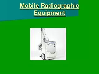
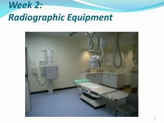
Radiographic equipment:. (1) X-ray tube- - anode/cathode- tube stand allows for sliding-longitudinal, transverse, and vertical.. (2) Collimator- below x-ray tube- controls size and shape of x-ray field- use a light source to project a representation of the x-ray field onto the patient

E N D
1. Radiographic and Fluoroscopy Equipment
2. Radiographic equipment: (1) X-ray tube-
- anode/cathode
- tube stand allows for sliding-longitudinal, transverse, and vertical.
4. (3) X-ray table- tilt, non-tilt, free floating or stationary and height adjustable or nonadjustable.
Tilt from horizontal to vertical
Bucky tray- below table, holds x-ray cassette, can be manually moved the entire length of the table and locked into place
6. console kVp- 1 kv=1000 volts
minor and major knobs �range 30-150 kvp
mA- 1 mA= 1/1000 of an ampere-
amt of current- range 100-400 mA for dx radiography
Time- seconds- 25% increments
mAs- how many x-rays for how long
Exposure- activate the rotor to begin- cause anode to rotate-prepare tube for exposure-second switch takes the exposure- automatic timer shuts it off.
7. Wall mounted Bucky Standing patients
Pull out tray and place cassette in the tray and lock it in place- can move entire Bucky system along vertical plane.
Alignment- table, tube and Bucky all move separately- be careful to check after positioning the patient that all parts are in alignment.
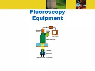
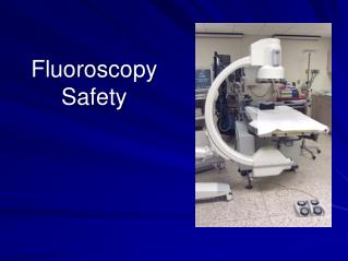
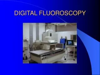
8. Components of Fluoroscopy X-ray tube- located under x-ray table
opposite from the tube-image intensifier- intercept attenuated beam-transform into electronic image.
Television monitor displays the image
Radiologist views a physiologic event