Download
1 / 72
810 likes | 1.22k Views
Brain Tumor Information Module. Basic Brain Information and Anatomy.

E N D
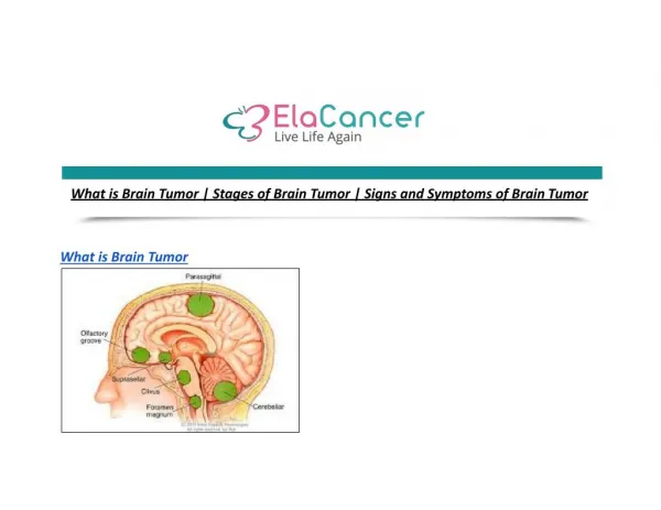
The brain is the major control center of a person’s body and mind. It is where ideas, senses, reflexes and movements originate. The human brain is extremely complex, but can be roughly organized into different structures that are responsible for different tasks.
Cerebral Cortex -The largest, most familiar part of the brain -Divided into left and right cerebral hemispheres that control the opposite sides of the body -responsible for reading, speech, thinking, learning, emotions, and reactions to senses. - Can be divided into more specific areas…
Divisions of the Cerebral Cortex • Frontal Lobe- gives you the ability to choose right from wrong, use correct social responses, and retain long term memories • Temporal Lobe – involved with speech, memory and hearing • Occipital Lobe – processes visual stimuli • Parietal Lobe – processes information to help with spatial orientation, manipulation of objects and understanding numbers
Inner Brain • The inner brain lies beneath the cerebral cortex and works as a connection between the cerebral cortex and the brainstem. Its structures determine our emotional state, consciousness, perceptions and reflexes.
Inner Brain Structures • Thalamus – processes information from the cortex and plays an important role in consciousness • Hypothalamus – Links the nervous system to the endocrine system, which makes it a major control center for emotions • Hippocampus – Responsible for memory formation and storage • Pituitary Gland – Controls hormone secretion
Inner Brain Structures • Basal Ganglia – A collection of nuclei that provide connections between the cortex, thalamus, and brainstem. These connections are involved with movement coordination, executing voluntary movements, perception, learning and memory. • Olfactory Bulb – Involved with smell. • Amygdala– Controls emotions, especially by forming emotional memories, and is also responsible for long-term memory.
Brain Stem • Responsible for essential life functions and relays information between the brain and the rest of the body • Midbrain – Controls reflexes and movement. • Pons - Responsible for breathing and arousal, and also coordinates movement information between the cortex and cerebellum. • Medulla – Maintains autonomic functions like breathing, blood pressure, and heart rate.
Cerebellum • Detects surroundings and coordinates movements to respond to specific situations
Important Links • Brain Anatomy (for fun) • 3-D Brain Anatomy • http://anatomyarcade.com/games/wordsearch/nervousWS/nervousWordsearch.html
Question • What are the 4 main lobes of the Cerebral Cortex? • What actions is the Cerebral Cortex responsible for?
Answer: • The Cerebral cortex is responsible for: • Frontal lobe • Long term memories, social interaction • Parietal lobe • Spatial orientation, object manipulation • Occipital lobe • Processing visual stimuli • Temporal lobe • Involved in speech, memory, and hearing
Meninges • The brain is a floating network of cells suspended in Cerebrospinal fluid (CSF). The CSF is produced by ependymal cells and circulates through the ventricles and meninges. The meninges cover the brain and attach it to the skull.
Meninges • We have 3 Meninges • Dura Mater • The tough layer next to the skull • Arachnoid Mater • The middle layer that is made up of web-like projections that connect the two other layers and allows CSF to flow through • Pia Mater • The thinnest layer that lies closely against the brain
Ventricles • There are four ventricles • The paired lateral ventricles • Third Ventricle • Fourth Ventricle • They are cavities in the brain that carry and promote the flow of cerebrospinal fluid
Question: • What are the three Meninges? • What flows through the Meninges • What are the big cavities in the brain that hold this fluid?
Answer • Dura Mater, Arachnoid Mater , Pia Mater • Cerebrospinal Fluid • Ventricles
Brain Matter • The brain is made up of neurons and glial cells. The neurons carry signals throughout the brain, while the glial cells mainly provide support for the neurons.
Neurons • 100 billion neurons in the brain • Have 3 basic parts • Dendrites • Detect signals from the surrounding cells and transmits them to the cell body • Cell body • Contains the nucleus, which is the control center for the cell • Axon • Carries signals from the cell body to the end of the axon • The axon is covered with myelin, which is an insulating sheath that helps to speed up signal movement
Glial Cells • Estimated 10x more glial cells than neurons • Do NOT chemo-electric carry signals • Main job is to hold neurons in place • Capable of secreting nutrients and other chemicals that can change signals sent throughout the brain
Types of Glial Cells Oligodendrocytes Schwann Cells • These cells produce myelin for the nerves outside of the brain and spinal cord • These cells produce myelin (insulating cover) for the nerves in the brain and spinal cord
Astrocytes Microglia Anchor to neurons and provide support Act as a buffer to absorb chemicals and promote homeostasis in the brain Eat foreign entities in the brain – part of the brain’s immune system
Ependymal Cells Line the ventricles in the brain Make CSF and helps it flow throughout the ventricles
Questions: • Are there more neurons or glial cells in the brain? • What is the main role of glial cells? • Specifically, what do Oligodendrocytes do? • Specifically, what do Astrocytes do?
Answers: • Glial cells – There is up to ten times more glial cells than neurons • Glial cells provide support • Oligodendrocytes make the insulating cover for neurons (myelin) • Astrocytes anchor neurons and absorb chemicals in the brain
Brain Tumor Information Brain Tumors arise when cell division occurs irregularly and uncontrolled. When more tissue is produced than needed, tumors are formed. They can grow in many different parts of the brain and involve different types of tissues and cells. If tumors are benign, they grow locally and do not spread. If a tumor is malignant, it is invasive and can spread throughout the body.
Malignant Brain Tumors • -When determining how malignant a brain tumor is, the cells are examined by a pathologist and given a grade. Brain tumors can be graded using the WHO classification system. • WHO Grade 1 – Well differentiated (Low Grade) • WHO Grade 2 – Moderately differentiated (Intermediate Grade) • WHO Grade 3 – Poorly Differentiated (High Grade) • WHO Grade 4 – Undifferentiated (High Grade)
Cell Differentiation Examples • A – Grade 2 • B – Grade 3 • C – Grade 4
Symptoms of a Brain Tumor By Location: • Frontal lobe • Weakness, personality changes, speech disturbances • Parietal lobe • Loss or changes in sensation, changes in vision • Temporal lobe • Seizures, difficulty understanding, difficulties with language • Occipital lobe • Changes in vision • Cerebellum • Abnormal eye movements, loss of coordination, changes in gait, hearing loss, vertigo, headaches, nausea, vomiting
Diagnosis • The process of detecting and treating a brain tumor can be extremely stressful and complicated. • Once a patient goes to see the doctor, they do a basic neurological exam. This exam tests many functions, such as eye movements, pupil reactions, reflexes, hearing, mental abilities, facial movements, and balance and coordination. • If these are abnormal, the doctor may schedule an MRI or a CT Scan
MRI – Magnetic Resonance Imaging • The patient lies on a table that slides into a tunnel with a magnetic field • During the scan, radio waves are sent to the head. The different cell types in the brain cause the waves to bend, which are recognized by a computer that forms a picture.
CT Scan – Computed Tomography • For a CT Scan, a person is injected with a dye and then lies on a table. A big donut shaped machine circles the head and sends x-ray waves through the brain to measure the amount of rays that are emitted back in the machine vs. the amount that are absorbed. By putting together all of the signals, a computer forms a picture of the brain.
Biopsy • Once it is determined that there is a growth in the brain, a biopsy is done in order to make an accurate diagnosis. If the tumor is in an accessible location, a patient may choose to have the whole tumor removed and then send a sample away for the biopsy. If the tumor is inaccessible, a tiny needle can be inserted into the brain to capture a small sample of tissue. Once the samples are attained, they are sent to a neuropatholigst.
Pathology • The tissue samples are either frozen or dried, then sliced into very thin sections. • The slices are mounted on to slides and examined with a microscope • First, the cell type that the tumor originated from is determined • Next, the growth rate of the tumor is assessed • Finally, the type and grade of the tumor is diagnosed
Pathology Report • When a patient is diagnosed, the details are put into a document called a pathology report. Physicians write them with the intent that other physicians will read them, so they are often complex and difficult to decipher. • Get picture
Pathology Report Contents • Main Sections • Personal Information • Name, date of birth, etc. • Clinical History • A brief description of a person’s medical situation • Gross Description • Describes how the tissue looks to the naked eye • Microscopic Description • Describes how the tissue looks under the microscope • Diagnosis • States the final Diagnosis • This is the biggest section of the report that specifically describes the type of tumor, what tissues are involved and patterns of growth. This section provides important information that is used to determine how to treat the patient’s specific tumor type. • Comments
Questions • What are some symptoms of brain tumors? • How is the level of malignancy graded? • What types of scans are done to see if a person has a brain tumor? • What document gives the results of a tumor analysis?
Answers: • Seizure, Nausea, Headaches, Weakness, Fatigue, Mood or Personality changes • WHO grade I-IV • MRI and CT Scan • Pathology report
Types of Brain Tumors • Some are from a single cell type • Ex.- Astrocytoma, Oligodendroglioma • Some are from a mixed cell type • Ex. – Oligoastrocytoma, mixed glioma • Some are mixed with neurons • Ex. – Ganglioma • Some are from neurons • Gangliocytoma • Some are from other tumors that have metastasized • About 40% of brain tumors are metastatic • The most common cancers that spread to the brain are lung, breast, melanoma, renal and colon cancers
Astrocytomas • Arise from Astrocyte cells - Astrocytoma Video • 4 types • PilocyticAstrocytoma (grade I) • Occurs mostly in children, benign • Low-Grade Astrocytoma (Grade II) • Can be removed by surgery, but radiation is also recommended • AnaplasticAstrocytoma (Grade III) • Radiation and Chemotherapy recommended • GlioblastomaMultiforme (Grade IV) • Very aggressive and spreads throughout the CNS • Patients usually have neurological symptoms • Radiation and chemotherapy recommended • Account for about 25% of all brain tumors • Life expectancy is about a year • GBM Video
Oligodendroglioma • Arise from oligodendrocytes • Average age of diagnosis = 35 years • 9.4% of all primary brain and CNS tumors • Occur most frequently in the frontal lobe • Primary symptom is usually a seizure • Usually grade II or III • Median survival • 11.6 years for grade II • 3.5 years for grade III • Usually grows slower than an Astrocytoma
Mixed Glioma • Most often a mix between an astrocytoma and an oligodendroglioma = oligoastrocytoma • Primarily occurs people aged 20 – 50 • Account for 1% of all brain tumors • Symptoms • Headache • Nausea and vomiting • Behavioral changes • Treatment based on most malignant cell type
Intraventricular Tumor • Make up about 10% of CNS tumors • May be composed of surrounding cells • Astrocytoma, Meningioma etc. • May arise from cells lining the ventricle • Ependymoma • 5% of CNS tumors • Survival rate of 5-10 years • 85% are benign • Tumor can block flow of Cerebrospinal fluid through the ventricles and cause obstructive hydrocephalus • May cause nausea, vomiting, deteriorating mental status, headache, neurological defects • Standard treatment includes surgery, and then radiation and/or chemotherapy if needed
Primary CNS Lymphoma • 90% are diffuse large B-cell lymphomas • Can also be poorly characterized low-grade lymphomas, Burkitt lymphomas, and T-cell lymphomas • Incidence increasing – especially among immunocompromised patients • Most commonly occurs around age 55 • Survival with radiation and chemotherapy is around 44 months
Metastatic Brain Tumors • Not a primary brain tumor • Composed of cancer cells that have spread from their original location • Most common: • Breast, Melanoma, Lung, Kidney • Treatment is composed of radiation and surgery if possible • Chemotherapy has not been found to be helpful • Surgery is not done when there are multiple tumors
Question • What is the name of a Grade IV astrocytoma? • What is the average life expectancy for a Grade IV astrocytoma? • Oligodendrogliomas are usually what Grades? • What is a type of Intraventricular Tumor?
Answer: • Glioblastoma Multiforme • 1 year • Grade II and III • Ependymoma