Download

1 / 38
380 likes | 536 Views
بنام خداي زيبائيها. Major Histocompatibility complex OR. MHC. MHC. The principal function of T cells are : Defense against intracellular microbes Activation of other cells(B cells ,Macrophages ) peptide recognition by T and B cells is different.

E N D
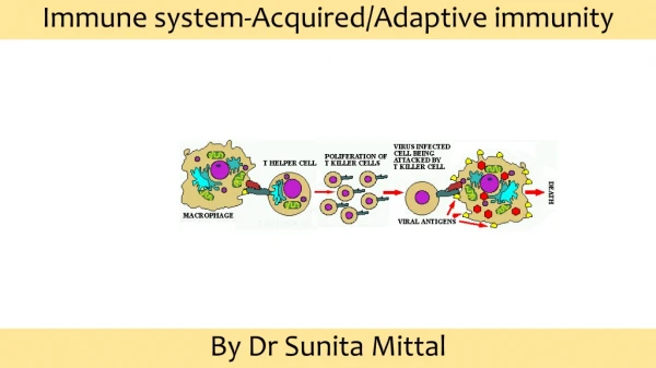
MHC • The principal function of T cells are : Defense against intracellular microbes Activation of other cells(B cells ,Macrophages ) • peptide recognition by T and B cells is different. B cells recognize soluble as well as cell associated Ags In contrast ,T cells recognize peptide which is displaying only by APC in associated with MHC proteins.
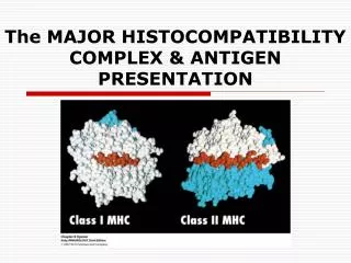
MHC • MHC genes are the most polymorphic genes • MHC genes are codominantly expressed in each individual • There are two main type of MHC:class I & II • The physiologic function of MHC molecules is presentation of peptides to T cells ,control of immune responsiveness to all proteins and graft rejection • MHC-I present cytosolic peptides to T cytotoxic cells and MHC-II present endocytosed peptides to Thcells
HLA-G ( a role in Ag recog.by NK cells) • HLA-H (involved in iron metabolism • HLA-DM (peptide binding to class II)
Class III • C4,B,C2 • TNF,LT joined to class II Proteasomegenes,TAP,DM
Conformational structure MHC-II MHC-I
Characteristics of peptide-MHC interaction • Each class –I or II has a single peptide binding cleft that are accommodate many different peptides • Slow on-rate and very slow off-rate • The MHC molecules of an individual don’t discriminate between self and non self
Polymorphism of class II • HLA-DPA 12 • HLA-DPB 88 • HLA-DQA 17 • HLA-DQB 42 • HLA-DR >400
Polymorphism of class I • HLA-A >280 • HLA-B >500 • HLA-C >130
Testing of DNA sequences permits detection of many more subtypes or "splits" of HLA antigens or alleles. • In serological typing, some antigens are difficult to identify and may even mask the presence of others. DNA typing can routinely define antigens at the allele level, assuring no ambiguity in interpretations. • DNA typing does not require live blood cells from the patient, permitting more flexible sample requirements. Thus, LabCorp can perform DNA-based HLA typing on:
Erythrocytes will adsorb some Class I antigens viz. Bg blood group system (B7,A28, B57….) HLA B most polymorphic system and studies have shown is most significant followed by A and then C 45Kd glycoprotein comprising three heavy chain domains, non-covalently associated More interesting facts
TYPING METHODS • SEROLOGY used to be the ‘gold’ standard. Now being superceded by molecular techniques as they become more robust and time efficient • CELLULAR rarely used now. Orginally used for Class II typing • MOLECULAR fast becoming the method of choice. Many laboratories test of choice.
SEROLOGY • Complement Dependent Cytotoxicity (CDC) • Viable peripheral blood lymphocytes are obtained by discontinous density gradient centrifugation using Ficoll / Tryosil or Ficoll / Sodium Metrizoate at a density of 1.077 at 19º - 22ºC. • Microlymphocytotoxic test: 3 stages
Microlymphocyototoxic test • 1.Viable lymphocytes are incubated with HLA specific antibodies. If the specific antigen is present on the cell the antibody is bound. • 2.Rabbit serum as a source of complement is added, incubate. If antibody is bound to the HLA antigen on the cell surface it activates the complement which damages the cell membrane making it permeable to vital stains.
Microlymphocyototoxic test 2 • 3.Results are visualised by adding dye usually a fluorochromeegEthidium Bromide although both Trypan Blue and Eosin have been used in the past. • If the reaction has taken place the EB enters the cell and binds to the DNA. • For ease double staining is normally used. We use a cocktail of Ethidium Bromide and Acridine Orange, quenched using Bovine Haemoglobin to allow simultaneous visualisation of both living and dead cells.
Microlymphocytotoxicity test 3 • Test is left for 10 minutes and then read using an inverted fluorescient microscope. • A mixture of T and B lymphocytes can be used for HLA Class I typing. • B lymphocytes are required for HLA Class II typing by serology. (Normal population 85-90% T and 10-15% B cells) • This can be achieved using a number of methods.
Easily performed does not require expensive equipment. • Takes around three hours to perform • Low level resolution, with good antisera reliable results • Requires large volumes of blood • Requires viable lymphocytes • Difficult to find good antisera for rarer antigens in different populations
molecular • DNA extraction from the nucleated cells following cell lysis and protein digestion. • polymerase chain reaction (PCR)
Molecular Methods 4 • Electrophoresis is used following amplification. PCR product is run out on an agarose gel containing ethidium bromide. Each product moves according to its size and is compared to a molecular weight marker. • Interpretation: every tube should produce an identical sized product as internal control and either a specific band or not dependent on whether the allele(s) is/are present or not. • Results are visualised using 312nm UV transillumination and recorded either by video imaging or polaroidphotograghy.