Download
1 / 33
330 likes | 581 Views
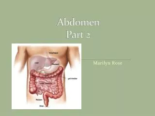
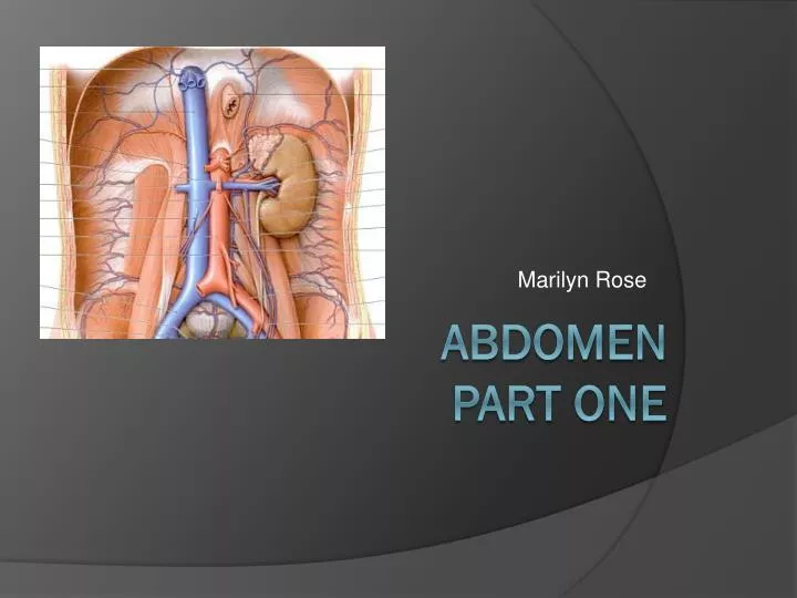
Marilyn Rose. Abdomen Part One. Abdominal cavity ***Between the diaphragm and sacral promontory*****. Peritoneum Thin serous membrane Parietal- lines abd walls Visceral- covers organs The cavity houses- liver (except bare area), GB, spleen, stomach, ovaries and most intestines

E N D
Marilyn Rose AbdomenPart One
Abdominal cavity ***Between the diaphragm and sacral promontory***** • Peritoneum • Thin serous membrane • Parietal- lines abd walls • Visceral- covers organs • The cavity houses- liver (except bare area), GB, spleen, stomach, ovaries and most intestines • Males- closed cavity • Females- communicates with exterior though fallopian tubes, UT, and vagina
Peritoneum contd. • Greater Sac communicates with the lesser Sac by the Foramen of Winslow (epiploic foramen) • Folds in the peritoneum= mesentery, omenta and peritoneal ligaments. • Mesentery encloses intestines and attaches them to the abd wall • Omentum- mesentery attaching to stomach • Greater- connects > curve of sto to spleen/ TRV colon • Lesser- connects duodenum, < curve to liver.
Ligaments • 3: > omental ligaments: • Gastrocolic, gastrosplenic and gastrophrenic • 2: < omental: • Hepatogastric, hepatoduodenal • 2: Liver ligaments: • Round (ligamentumteres) • Falciform • Diaphragm to liver: • Coronary ligaments • Margins of the “bare area”
Bare Area of the Liver The coronary ligaments represent reflections of the visceral peritoneum covering the liver onto the diaphragm. As such, between the two layers of the coronary ligament there is a large triangular surface of the liver devoid of peritoneal covering; this is named the bare area of the liver, and is attached to the diaphragm by areolar tissue.
Peritoneal spaces • Supracolic compartment • Above transverse colon • R/L subphrenic spaces • Between diaph and anterior liver- R/L by falciform ligament • R/L subhepatic spaces • Post/ inferior btw liver and abdominal viscera • Rt= Morison’s Pouch– deepest in supine pt and common site for fluid collections!!
Peritoneal spaces • Infracolic compartment • Below transverse colon • R/L infracolic spaces • Divided by mesentery of small intestine • paracolic gutters • Lateral to ascentind and descending colon • Deeper RT is also a common site for fluid collections….
Peritoneal Spaces What is the inflammation of the peritoneum and what is the most common cause???
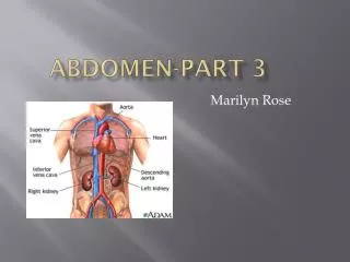
Retroperitoneum • Structures located posterior to peritoneum and still lined by it. • Kidneys/ ureters • Adrenal glands • Pancreas • Duodenum/ ascending/descending colon • AO • Inferior vena cava • Bladder • Uterus • Prostate gland
Retroperitoneal Spaces • Anterior pararenal space • Between Grota’s fascia/ post peritoneum • Ascending/descending colon and pancreas and duodenum. • Posterior pararenal space • Posterior renal fascia and muscles of posterior abd wall (only fat and vessels in this space) • LT/RT perirenal space areas directly around kidney • This space contains the kidneys, adrenal glands, lymph nodes, blood vessels and perirenal fat
Abdominal Aorta • Retroperitoneal • Delivers O2 blood to abdominopelvic structures • L4- bifurcates- Rt/Lt- Common iliac arteries
Abdominal Aorta • Celiac Trunk • Off anterior wall- 3 arteries • 1. left gastric • 2. common hepatic- (divides into the hepatic artery and gastroduodenal artery) • 3. splenic • SMA • L1- inferior to celiac and descends behind pancreas • Renal arteries • Arise from lateral walls of AO • Below SMA • RRA longer than left and passes posterior to IVC FYI: Renal artery stenosis can lead to HTN!!!!!
Normal Abnormal
Inferior Vena Cava • Largest vein in the body • Carries blood to the heart from the lower limbs, pelvic organs and abdomen • L5- is formed by the common iliac veins • Ascends superiorly through the retroperitoneum along the anterior verterbal column to the RT of the AO. • Renal veins- empty into IVC @ L2 • Lt renal vein- posterior to SMA and anterior to the aorta with the LT gonadal vein draining into it • Hepatic veins • 3 – Rt/ Middle/ Lt • Collects blood from liver parenchyma • Drain from inferior liver to the superior where they empty into the IVC below the diaphragm and proximal to the Rt atrium
TIPS- RA-stent
If the IVC thrombosis…what then? AZYGOUS RECANALIZATION
Biliary Atresia… Leads to cirrhosis & MPV thrombosis
Hepatoblastoma How do we fix these??