Download
1 / 32
320 likes | 532 Views
. 1. Origine inflammatoire . Sclrose en plaques . Affection neurologique inflammatoire, volutive 2-6 nouveaux cas/an/100 000 Fe 20-30 ans - Ho 30- 40 ans tiologie ? processus auto-immun / perturbation du systme immunitaire / environnement / susceptibilit gntique/. cliniques para

E N D
7. BaloBalo
15.
18.
NB. Pas de corr�lations entre clinique et HS => comment expliquer ? Une des explications est que la technique n�est pas adapt�e. Ici, le 3D FLAIR et mieux le 3D DIR => Correct determination of cortical-subcortical lesion load is imperative for valid clinical and neuropsychologic studies in patients with multiple sclerosis. Results of previous studies in which FLAIR MR was used to determine (juxta)cortical lesion count or load (14,16,36) have shown poor correlations with clinical disability. Accurate cortical lesion detection by use of 3D DIR imaging should be a very helpful tool in understanding more about the physical and cognitive problems encountered by patients with multiple sclerosis. The present study did not specifically focus on correlating lesion counts with clinical and neuropsychologic measures but merely showed the possible improvement in detection of intracortical lesions. It is therefore essential that future studies focus on correlating well-defined cortical-subcortical lesion load to specific measures of disability and neuropsychologic function in a larger group of patients. Moreover, follow-up studies performed to evaluate changes in cognitive functioning in relation to increasing cortical disease burden will provide useful and important information. In conclusion, 3D DIR MR imaging shows increased depiction of intracortical lesions in brains with multiple sclerosis, as well as increased definition when assessing mixed white matter�gray matter lesions.
Geurts JJ et al. AJNR Am J Neuroradiol. 2005;26:572-7. BACKGROUND AND PURPOSE: Cortical lesions constitute a substantial part of the total lesion load in multiple sclerosis (MS) brain. They have been related to neuropsychological deficits, epilepsy, and depression. However, the proportion of purely cortical lesions visible on MR images is unknown. The aim of this study was to determine the proportion of intracortical and mixed gray matter (GM)-white matter (WM) lesions that can be visualized with postmortem MR imaging. METHODS: We studied 49 brain samples from nine cases of chronic MS. Tissue sections were matched to dual-echo T2-weighted spin-echo (T2SE) MR images. MS lesions were identified by means of myelin basic protein immunostaining, and lesions were classified as intracortical, mixed GM-WM, deep GM, or WM. Investigators blinded to the histopathologic results scored postmortem T2SE and 3D fluid-attenuated inversion recovery (FLAIR) images. RESULTS: Immunohistochemistry confirmed 70 WM, eight deep GM, 27 mixed GM-WM, and 63 purely cortical lesions. T2SE images depicted only 3% of the intracortical lesions, and 3D FLAIR imaging showed 5%. Mixed GM-WM lesions were most frequently detectable on T2SE and 3D FLAIR images (22% and 41%, respectively). T2SE imaging showed 13% of deep GM lesions versus 38% on 3D FLAIR. T2SE images depicted 63% of the WM lesions, whereas 3D FLAIR images depicted 71%. Even after side-by-side review of the MR imaging and histopathologic results, many of the intracortical lesions could not be identified retrospectively. CONCLUSION: In contrast to WM lesions and mixed GM-WM lesions, intracortical lesions remain largely undetected with current MR imaging resolution.
21. ADEM : IRM
24. Observation Mme G. 51 ans : �thylisme chronique (bi�re) d�pression
d�nutrition, asth�nie, vomissement, d�shydratation extracellulaire
examen neurologique normal
Na+ : 118 mmol/l K+ : 2,4 mmol/l
26. Atteinte �lective du tronc c�r�bral 1. Infection : listeriose
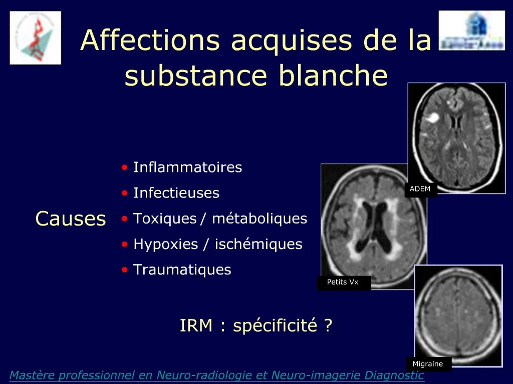
Contre : pas de prise de contraste
2. Maladie de syst�me : Beh�et
3. Tumeur infiltrante
Contre : Contraste, effet de masse -
4. SEP, ADEM
Contre : pas multifocal
5. Isch�mie art�rielle
Contre : pauci-symptomatique