Download
1 / 27
390 likes | 920 Views
Gram-Negative Outer Membrane. Figure 4.13c. Figure 4.13b–c. Thick peptidoglycan Teichoic acids In acid-fast cells, contains mycolic acid. Thin peptidoglycan No teichoic acids Outer membrane. Gram-Positive Gram-Negative Cell Walls Cell Walls. Gram-Positive Cell Walls. Teichoic acids

E N D
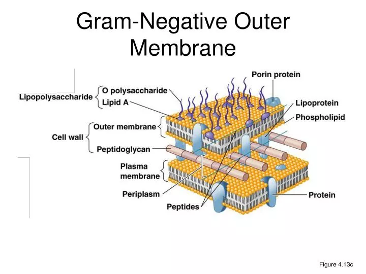
Gram-Negative Outer Membrane Figure 4.13c
Thick peptidoglycan Teichoic acids In acid-fast cells, contains mycolic acid Thin peptidoglycan No teichoic acids Outer membrane Gram-Positive Gram-Negative Cell Walls Cell Walls
Gram-Positive Cell Walls • Teichoic acids • Lipoteichoic acid links to plasma membrane • Wall teichoic acid links to peptidoglycan • May regulate movement of cations. • Polysaccharides provide antigenic variation. Figure 4.13b
Gram-Negative Outer Membrane • Lipopolysaccharides, lipoproteins, phospholipids • Forms the periplasm between the outer membrane and the plasma membrane. • Protection from phagocytes, complement, and antibiotics • O polysaccharide antigen, e.g., E. coli O157:H7 • Lipid A is an endotoxin • Porins (proteins) form channels through membrane.
Gram Stain Mechanism • Crystal violet-iodine crystals form in cell. • Gram-positive • Alcohol dehydrates peptidoglycan • CV-I crystals do not leave • The color will be purple • Gram-negative • Alcohol dissolves outer membrane and leaves holes in peptidoglycan. • CV-I washes out • The color will be purple
Atypical Cell Walls • Mycoplasmas • Lack cell walls • Sterols in plasma membrane • Archaea • Wall-less or • Walls of pseudomurein (lack NAM and D amino acids)
Damage to Cell Walls • Lysozyme digests disaccharide in peptidoglycan. • Penicillin inhibits peptide bridges in peptidoglycan. • Protoplast is a wall-less cell. • Spheroplast is a wall-less Gram-positive cell. • L forms are wall-less cells that swell into irregular shapes. • Protoplasts and spheroplasts are susceptible to osmotic lysis.
Plasma Membrane Figure 4.14a
Plasma Membrane • Phospholipid bilayer • Peripheral proteins • Integral proteins • Transmembrane proteins Figure 4.14b
Fluid Mosaic Model • Membrane is as viscous as olive oil. • Proteins move to function. • Phospholipids rotate and move laterally. Figure 4.14b
Plasma Membrane • Selective permeability allows passage of some molecules • Enzymes for ATP production • Photosynthetic pigments on foldings called chromatophores or thylakoids
Plasma Membrane • Damage to the membrane by alcohols, quaternary ammonium (detergents), and polymyxin antibiotics causes leakage of cell contents.
Movement Across Membranes • Simple diffusion: Movement of a solute from an area of high concentration to an area of low concentration. • Facilitative diffusion: Solute combines with a transporter protein in the membrane.
Movement Across Membranes Figure 4.17
Movement Across Membranes • Osmosis: The movement of water across a selectively permeable membrane from an area of high water concentration to an area of lower water. • Osmotic pressure: The pressure needed to stop the movement of water across the membrane. Figure 4.18a
Movement Across Membranes Figure 4.18a–b
Movement Across Membranes • Active transport of substances requires a transporter protein and ATP. • Group translocation of substances requires a transporter protein and PEP.
Cytoplasm • Cytoplasm is the substance inside the plasma membrane. Figure 4.6a–b
Nuclear Area • Nuclear area (nucleoid) Figure 4.6a–b
Ribosomes Figure 4.6a–b
Ribosomes Figure 4.19
Inclusions Figure 4.20
Metachromatic granules (volutin) Polysaccharide granules Lipid inclusions Sulfur granules Carboxysomes Gas vacuoles Magnetosomes Phosphate reserves Energy reserves Energy reserves Energy reserves Ribulose 1,5-diphosphate carboxylase for CO2 fixation Protein covered cylinders Iron oxide (destroys H2O2) Inclusions
Endospores • Resting cells • Resistant to desiccation, heat, chemicals • Bacillus, Clostridium • Sporulation: Endospore formation • Germination: Return to vegetative state