Download
1 / 56
590 likes | 775 Views
The Plasma Membrane Biology 11 Version II E. McIntyre. History of the Plasma Membrane. 1665: Robert Hooke 1895: Charles Overton - composed of lipids 1900-1920’s: must be a phospholipid 1925: E. Gorter and G. Grendel - phospholipid bilayer

E N D
History of the Plasma Membrane • 1665: Robert Hooke • 1895: Charles Overton - composed of lipids • 1900-1920’s: must be a phospholipid • 1925: E. Gorter and G. Grendel - phospholipid bilayer • 1935: J.R. Danielli and H. Davson – proteins also part, proposed the Sandwich Model • 1950’s: J.D. Robertson – proposed the Unit Membrane Model • 1972: S.J. Singer and G.L. Nicolson – proposed Fluid Mosaic Model
Plasma Membrane is made of Phospholipids • Gorter + Grendel • Red Blood Cells analyzed • Enough for Phospholipid bilayer • Polar heads face out and Nonpolar tails face in • Does not explain why some nonlipids are permeable
Plasma Membrane Models Sandwich Model (Danielli + Davson) 2 layers of globular proteins with phospholipid inside to make a layer and then join 2 layers together to make a channel for molecules to pass Unit Membrane Model (Robertson) Outer layer of protein with phospholipid bilayer inside, believed all cells same composition, does not explain how some molecules pass through or the use of proteins with nonpolar parts, used transmission electron microscopy Fluid Mosaic Model (Singer + Nicolson) Phospholipid bilayer with proteins partially or fully imbedded, electron micrographs of freeze-fractured membrane
Which membrane model is correct? 1) Rapidly freeze specimen 2) Use special knife to cut membrane in half 3) Apply a carbon + platinum coating to the surface 4) Use scanning electron microscope to see the surface According to the electron micrograph which membrane model is correct? Why? Fluid-Mosaic Model
Fluid-Mosaic Model • Fluid – the plasma membrane is the consistency of olive oil at body temperature, due to unsaturated phospholipids. (cells differ in the amount of unsaturated to saturated fatty acid tails) • Most of the lipids and some proteins drift laterally on either side. Phospholipids do not switch from one layer to the next. • Cholesterol affects fluidity: at body temperature it lessens fluidity by restraining the movement of phospholipids, at colder temperatures it adds fluidity by not allowing phospholipids to pack close together. • Mosaic – membrane proteins form a collage that differs on either side of the membrane and from cell to cell (greater than 50 types of proteins), proteins span the membrane with hydrophilic portions facing out and hydrophobic portions facing in. Provides the functions of the membrane
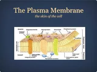
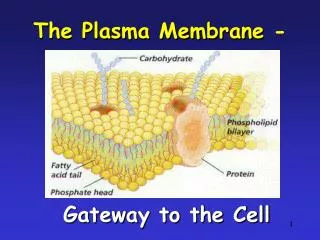
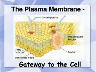
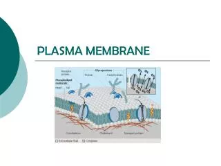
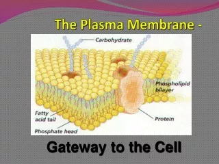
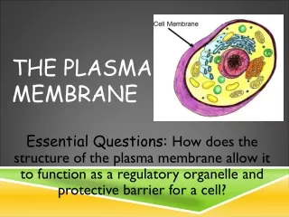
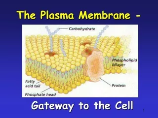
Structure of the Plasma Membrane • Phospholipid bilayer • Phospholipid • Hydrophilic head • Hydrophobic tails • Cholesterol • Proteins • Transmembrane/ Intrinsic/Integral • Peripheral/Extrinsic • Cytoskeletal filaments • Carbohydrate chain • Glycoproteins • Glycolipids
Label the Blank Diagram of the Plasma Membrane • Phospholipid bilayer • Phospholipid • Hydrophilic head • Hydrophobic tails • Cholesterol • Proteins • Transmembrane/ Intrinsic/Integral • Peripheral/Extrinsic • Cytoskeletal filaments • Carbohydrate chain • Glycoproteins • Glycolipids
Proteins of the Plasma Membrane Provide 6 Membrane Functions: 1) Transport Proteins 2) Receptor Proteins 3) Enzymatic Proteins 4) Cell Recognition Proteins 5) Attachment Proteins 6) Intercellular Junction Proteins
1)Transport Proteins Channel Proteins – channel for lipid insoluble molecules and ions to pass freely through Carrier Proteins – bind to a substance and carry it across membrane, change shape in process
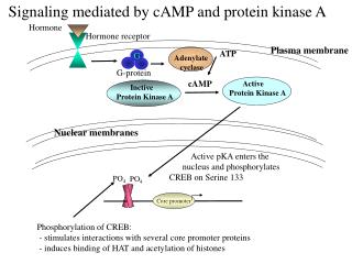
2)Receptor Proteins – Bind to chemical messengers (Ex. hormones) which sends a message into the cell causing cellular reaction
3)Enzymatic Proteins – Carry out enzymatic reactions right at the membrane when a substrate binds to the active site
4)Cell Recognition Proteins – Glycoproteins (and glycolipids) on extracellular surface serve as ID tags (which species, type of cell, individual). Carbohydrates are short branched chains of less than 15 sugars
5)Attachment Proteins • Attach to cytoskeleton (to maintain cell shape and stabilize proteins) and/or the extracellular matrix (integrins connect to both). • Extracellular Matrix – protein fibers and carbohydrates secreted by cells and fills the spaces between cells and supports cells in a tissue. • Extracellular matrix can influence activity inside the cell and coordinate the behavior of all the cells in a tissue.
6)Intercellular Junction Proteins – Bind cells together • Tight junctions • Gap junctions
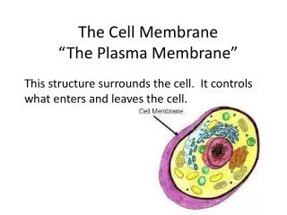
How do materials move into and out of the cell? • Materials must move in and out of the cell through the plasma membrane. • Some materials move between the phospholipids. • Some materials move through the proteins.
Plasma Membrane Transport • Molecules move across the plasma membrane by: Active Transport Passive Transport
What are three types of passive transport? • Diffusion • Facilitated Diffusion • Osmosis Passive Transport ATP energy is not needed to move the molecules through.
Passive Transport 1: Diffusion • Molecules can move directly through the phospholipids of the plasma membrane This is called … DIFFUSION
What is Diffusion? • Diffusion is the net movement of molecules from a high concentration to a low concentration until equally distributed. • Diffusion rate is related to temperature, pressure, state of matter, size of concentration gradient, and surface area of membrane. http://www.biologycorner.com/resources/diffusion-animated.gif
What molecules pass through the plasma membrane by diffusion? • Gases (oxygen, carbon dioxide) • Water molecules (rate slow due to polarity) • Lipids (steroid hormones) • Lipid soluble molecules (hydrocarbons, alcohols, some vitamins) • Small noncharged molecules (NH3)
Why is diffusion important to cells and humans? • Cell respiration • Alveoli of lungs • Capillaries • Red Blood Cells • Medications: time-release capsules
Passive Transport 2: Facilitated Diffusion • Molecules can move through the plasma membrane with the aid of transport proteins This is called … FACILITATED DIFFUSION
What is Facilitated Diffusion? • Facilitated diffusion is the net movement of molecules from a high concentration to a low concentration with the aid of channel or carrier proteins.
What molecules move through the plasma membrane by facilitated diffusion? • Ions (Na+, K+, Cl-) • Sugars (Glucose) • Amino Acids • Small water soluble molecules • Water (faster rate)
How do molecules move through the plasma membrane by facilitated diffusion? • Channel and Carrier proteins are specific: • Channel Proteins allow ions, small solutes, and water to pass • Carrier Proteins move glucose and amino acids • Facilitated diffusion is rate limited, by the number of proteins channels/carriers present in the membrane.
Specific Types of Facilitated Diffusion • Counter Transport – the transport of two substances at the same time in opposite directions, without ATP. Protein carriers are called Antiports. • Co-transport – the transport of two substances at the same time in the same direction, without ATP. Protein carriers are called Symports. • Gated Channels – receptors combined with channel proteins. When a chemical messenger binds to a receptor, a gate opens to allow ions to flow through the channel.
Why is facilitated diffusion important to cells and humans? • Cells obtain food for cell respiration • Neurons communicate • Small intestine cells transport food to bloodstream • Muscle cells contract
Passive Transport 3: Osmosis • Water Molecules can move directly through the phospholipids of the plasma membrane This is called … OSMOSIS
What is Osmosis? • Osmosis is the diffusion of water through a semipermeable membrane. Water molecules bound to solutes cannot pass due to size, only unbound molecules. Free water molecules collide, bump into the membrane, and pass through.
Osmosis in action • What will happen in the U-tube if water freely moves through the membrane but glucose can not pass? • Water moves from side with high concentration of water to side with lower concentration of water. Movement stops when osmotic pressure equals hydrostatic pressure.
Why is osmosis important to cells and humans? • Cells remove water produced by cell respiration. • Large intestine cells transport water to bloodstream • Kidney cells form urine
Osmosis and Tonicity • Tonicity refers to the total solute concentration of the solution outside the cell. • What are the three types of tonicity? • Isotonic • Hypotonic • Hypertonic
Isotonic • Solutions that have the same concentration of solutes as the suspended cell. • What will happen to a cell placed in an Isotonic solution? • The cell will have no net movement of water and will stay the same size. • Ex. Blood plasma has high concentration of albumin molecules to make it isotonic to tissues.
Hypotonic • Solutions that have a lower solute concentration than the suspended cell. • What will happen to a cell placed in a Hypotonic solution? • The cell will gain water and swell. • If the cell bursts, then we call this lysis. (Red blood cells = hemolysis) • In plant cells with rigid cell walls, this creates turgor pressure.
Hypertonic • Solutions that have a higher solute concentration than a suspended cell. • What will happen to a cell placed in a Hypertonic solution? • The cell will lose water and shrink. (Red blood cells = crenation) • In plant cells, the central vacuole will shrink and the plasma membrane will pull away from the cell wall causing the cytoplasm to shrink called plasmolysis.
Review: Passive Transport • Diffusion – O2 moves in and CO2 moves out during cell respiration • Facilitated Diffusion – glucose and amino acids enter cell for cell respiration • Osmosis – cell removal or addition of water
Review Tonicity • What will happen to a red blood cell in a hypertonic solution? • What will happen to a red blood cell in an isotonic solution? • What will happen to a red blood cell in a hypotonic solution?
What are three types of Active transport? 1) Active Transport 2) Exocytosis 3) Endocytosis • Phagocytosis • Pinocytosis • Receptor-Mediated endocytosis Active Transport ATP energy is required to move the molecules through.
Active Transport • Molecules move from areas of low concentration to areas of high concentration with the aid of ATP energy. • Requires protein carriers called Pumps.
The Importance of Active Transport • Bring in essential molecules: ions, amino acids, glucose, nucleotides • Rid cell of unwanted molecules (Ex. sodium from urine in kidneys) • Maintain internal conditions different from the environment • Regulate the volume of cells by controlling osmotic potential • Control cellular pH • Re-establish concentration gradients to run facilitated diffusion. (Ex. Sodium-Potassium pump and Proton pumps)
The Sodium-Potassium Pump • 3 Sodium ions move out of the cell and then 2 Potassium ions move into the cell. • Driven by the splitting of ATP to provide energy and conformational change to proteins by adding and then taking away a phosphate group. • Used to establish an electrochemical gradient across neuron cell membranes. http://www.biologie.uni-hamburg.de/b-online/library/biology107/bi107vc/fa99/terry/images/ATPpumA.gif
Active Transport 2: Exocytosis • Movement of large molecules bound in vesicles out of the cell with the aid of ATP energy. Vesicle fuses with the plasma membrane to eject macromolecules. • Ex. Proteins, polysaccharides, polynucleotides, whole cells, hormones, mucus, neurotransmitters, waste
Active Transport 3: Endocytosis • Movement of large molecules into the cell by engulfing them in vesicles, using ATP energy. • Three types of Endocytosis: • Phagocytosis • Pinocytosis • Receptor-mediated endocytosis
Phagocytosis • “Cellular Eating” – engulfing large molecules, whole cells, bacteria • Ex. Macrophages ingesting bacteria or worn out red blood cells. • Ex. Unicellular organisms engulfing food particles.
Pinocytosis • “Cellular Drinking” – engulfing liquids and small molecules dissolved in liquids; unspecific what enters. • Ex. Intestinal cells, Kidney cells, Plant root cells
Receptor-Mediated Endocytosis • Movement of very specific molecules into the cell with the use of vesicles coated with the protein clathrin. • Coated pits are specific locations coated with clathrin and receptors. When specific molecules (ligands) bind to the receptors, then this stimulates the molecules to be engulfed into a coated vesicle. • Ex. Uptake of cholesterol (LDL) by animal cells
Review Types of Endocytosis • What is phagocytosis? • What is pinocytosis? • What is receptor-mediated endocytosis?
In Animal Cells: Tight Junctions Desmosomes Gap Junctions In Plant Cells: Plasmodesmata Types of Cell Junctions