Download
1 / 85
1.21k likes | 2.8k Views
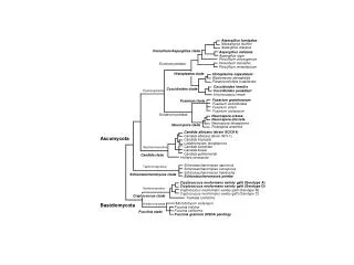
Phylum - Ascomycota. Kingdom - Fungi. Higher fungi. Two phyla – Ascomycota and Basidiomycota Thallus in both consists of sepatate hyphae that form extensive mycelia Septa have pores that allow migration of cytoplasm, organelles and nuclei (specialized septa in Basidiomycota)

E N D
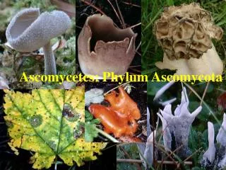
Phylum - Ascomycota Kingdom - Fungi
Higher fungi • Two phyla – Ascomycota and Basidiomycota • Thallus in both consists of sepatate hyphae that form extensive mycelia • Septa have pores that allow migration of cytoplasm, organelles and nuclei (specialized septa in Basidiomycota) • Compartments contain multiple nuclei
Higher fungi • Vegetative hyphae can fuse with one another = anastomosis (does not occur in lower fungi) • Asexual reproduction by conidia on conidiophores
Higher fungi • In sexual cycle, plasmogamy is separated from karyogamy – produces dikaryotic phase • Sexual reproduction produces spores after meiosis – ascospores or basidiospores (1n)
Ascomycota • General characterisitics • Generally have a similar sexual life cycle • All produce an ascus (sac-like structure) that contains haploid (1n) ascospores after meiosis • Includes ~75% of the described fungi – 30,000 spp.+ 20,000 anamorphs
Ascomycota • Sexual life cycle is basically similar – haploid-dikaryotic life cycle • Vegetative phase is haploid mycelium that may reproduce asexually by formation of conidia • Plasmogamy is separated from karyogamy in time so that a dikaryotic phase is produced – the ascogenous hyphae represent the dikaryotic hyphae
Gametangia • Male gametangia may be an antheridium or conidium-like structure – spermatium • Female gametangium = ascogonium, may have a long projection, the trichogyne
Plasmogamy • Male element fuses with trichogyne of the ascogonium, nuclei migrate into the ascogonium to begin the dikaryotic condition • After plasmogamy, two hyphal systems begin to grow • Sterile haploid hyphae envelope the ascogonium to form the multicellular ascoma (fruiting body) • Dikaryotic ascogenous hyphae grow from ascogonium – will give rise to the asci
Ascogenous hyphae • Male and female nuclei divide conjugately to maintain the dikaryotic condition • Many ascogenous hyphae produced inside the developing ascoma • Tips of ascogenous hyphae form croziers (hooks) before developing into an ascus
Developing ascoma • Ascoma contains two types of hyphae • Ascogenous hyphae – dikaryotic, form asci through crozier formation • Sterile hyphae – haploid, form bulk of ascoma
Crozier and ascus formation • During ascus formation, karyogamy occurs to form diploid nucleus followed by meiosis to form 4 haploid nuclei Mitosis Meiosis Karyogamy
Ascus formation • Most asci are cylindrical, but may be globose • 8 ascospores/ascus is a common number but this may vary • In most, ascogenous hyphae continue to proliferate, forming more croziers and more asci
Types of asci • Great deal of variation in asci and ascospores • Three basic types of asci • Prototunicate asci – thin, delicate wall that deliqueses to release ascospores
Types of asci • Unitunicate asci – ascus wall layers adhere closely to one another, ascospores released through a pore, a slit or an operculum (hinged cap) • Operculate asci • Inoperculate asci
Types of asci • Bitunicate asci – two wall layers that separate with the inner wall expanding, ascospores released through a pore
Fruiting bodies - ascomata • In most, the ascogenous hyphae are produced only in the ascoma (pl. ascomata • Ascomata consist of two types of hyphae – dikaryotic ascogenous hyphae that form the asci and haploid sterile hyphae that form the bulk of the ascoma • Four major types of ascomata
Cleistothecium • The cleistothecium remain closed until broken by internal forces, the asci are produced randomly within the ascoma
Perithecium • Begins as a closed structure but produces a pore at maturity through which the ascospores can escape • Asci produced in a definite layer - hymenium
Apothecium • Ascoma is open when asci mature, asci are produced in an hymenium
Ascostroma • Asci are produced in a cavity (locule) within a mass of sterile tissue = stroma • No ascoma wall as stroma did not originate from ascogonium
Ascomycota classification • Currently in a state of flux • Most current treatments do not divide into classes, but rather orders • 43 (35) orders have been proposed in the Ascomycota, some of these only occur in lichens
Ascomycota classification • We will examine these fungi as: • Cleistothecial Ascomycota • Perithecial Ascomycota • Apothecial Ascomycota • Pseudothecial Ascomycota • Non ascomatal Ascomycota - yeasts • Will discuss representatives within each of these groups to get an introduction to the Ascomycota
Cleistothecial Ascomycota • Cleistothecia are the simplest type of ascoma • Surrounded by relatively simple tissue (loose hyphae), do not have an opening, are broken open • Asci are scattered • Asci are prototunicate, globose to pear shaped
Cleistothecial Ascomycota • Include species that grow as saprotrophs on keratin (protein in hair, nails) • Contains the teleomorphs of human pathogens • Dermatophytes – cause superficial skin infections such as atheletes foot • Anamorphs are Trichophyton and Microsporium • Also contains fungi that cause deep or systemic infections of humans • Ajellomyces, the teleomorph of Histoplasma capsulatum which causes histoplasmosis • Blastomyces which causes blastomycosis
Aspergillus & Penicillium • The anamorphic stages (Aspergillus and Penicillium) – better known than the teleomorphs • Asci are spherical to club shaped, ascus wall dissolves at maturity leaving ascospores free inside the cleistothecium • Most are saprotrophs • Widespread in soil, litter • Opportunistic human & animal pathogens – aspergilloses, penicilloses
Aspergillus & Penicillium • Important industrial organisms, used in making – • Chemicals – citric, gluconic & other organic acids • Antibiotics – penicillin, griseofulvin • Production of miso and soy sauce, sake • Cheese production – blue cheese and camembert • Also important in food spoilage • Citrus fruits • Grains & peanuts – produce mycotoxins
Aspergillus • Anamorphic genus – close to 100 species • 11 different teleomorphic genera produce Aspergillus conidia and conidiophores, including Eurotium, Emericella • Common fungi found in air, soil, water • Grow on a variety of substrates, in humid climates found growing on clothing, shoes, etc. • Important as contaminants of stored grain, species produce aflatoxin
Aspergillus • Produce characteristic conidiophore • Conidia produced by phialides – flask shaped conidiogenous cells • Have a characterisitic foot cell
Penicillium • Over 95 species connected to 3 teleomorphic genera – Talaromyces, Eupenicillium, Carpentales • Very common in soil, conidia found in air, water, soil • Food spoilage – on citrus fruits, jelly, cheeses • Produce penicillin and other chemicals industrially • P. roqfertii, P. camembertii used to make cheeses
Penicillium • Asexual conidiophore – not swollen at tip, no foot cell • Phialides arranged in a brushlike manner
Powdery mildews • Ascomata do not form opening so can be termed cleistothecia, but are more like perithecia without an opening • Asci occur in an irregular layer, not scattered • Asci are unitunicate, forcibly discharge ascospores through a slit • Produce colorless hyphae on surface of plant host • Produce haustoria that penetrate epidermal cells of host
Asexual reproduction • Hyphae produce chains of conidia during growing season (spring and summer) on surface of plant leaf – giving the powdery, white appearance
Sexual reproduction • Late in growing season, as plants begin to senesce, ascomata are produced – thought to overwinter as ascomata • Ascomata are closed, have characteristic appendages extending from them • Asci are globose to ovoid, generally one to a few asci/ascoma
Ascoma • Appendages
Perithecial Ascomycota • Ascoma is a perithecium • Has an opening through which ascospores leave the ascoma = ostiole • Asci produced in a layer = hymenium • Asci are unitunicate and inoperculate • Asci typically have a pore or slit at the thickened tip
Perithecial Ascomycota • A large and diverse group of fungi –(have already discussed the powdery mildews which are sometimes included here • Include species which are • Saprotrophs – wood, dung, soil, plant litter • Parasites – plant, animal (arthropod) diseases • Endophytes • Some produce mycotoxins
Perithecia • Typically flask shaped structures • Pore with opening = ostiole • Perithecial wall composed of pseudoparenchyma tissue • Centrum is the central part where asci develop
Perithecia • Perithecial wall composed of fungal tissue called pseudoparenchyma – thin cell walls, looks like plant parenchyma tissue • Centrum – the asci and sterile structures that fill the cavity within the perithecial wall • Sterile structures: • Paraphyses – basally produced in hymenium • Pseudoparaphyses – originate from top of the perithecium, grow into hymenium • Periphyses – extending into the ostiole
Perithecium Ostiole Periphysis • Classification is based in large part on development of centrum, development of paraphyses or pseudoparaphyses Wall Stroma Paraphysis Ascus Ascospore
Perithecia • Perithecia may be produced individually or they may be produced within a mass of tissue = stroma (always produce a separate perithecial wall)
Stroma (stromata) • Compact tissue that forms a flat plate or a mass • Hyphae may be inflated, intertwined, have lost identity to form a tissue that looks like parenchyma tissue of plants – called pseudoparenchyma • May be hard and woody or soft and fleshy
Perithecial Ascomycota • Saprotrophic fungi on dung, wood, in soil & decaying leaves • Includes Neurospora – important in study of genetics • Asci are club shaped to cylindrical, most produce 8 ascospores • Asci may breakdown and release ascospores or persist & forcibly discharge ascospores
Chaetomium • Common saprotrophs in soil and dung, highly cellulolytic • Perithecia have distinctive filaments (straight or spiral )extending from upper part of perithecium • Asci are club shaped and wall deliqueses leaving ascospores embedded in jelly like substance that oozes out of the ostiole
Chaetomium • Ascospores are lemon shaped
Neurospora crassa • Red (pink) bread mold – grows on dough in bakeries, forms lots of conidia, bad contaminant • Widely used in genetic studies, one gene – one enzyme concept developed in Neurospora • Mostspecies are heterothallic – two mating types – A & a
Ascospore discharge • In many Ascomycota, asci develop high turgor pressure and actively discharge ascospores individually or as a group from ascus • In some, particularly coprophilous forms, the asci or ascomata may be phototrophic so that ascospores are discharged toward the light
Ascospore discharge • In Sordaria, Neurospora and other members of this group, asci stretch through ostiole and actively discharge ascospores
Xylaria • Produce dark colored, brittle solitary perithecia or perithecia may be produced in a stroma • Stromata are hard and woody, generally dark colored • In development of centrum, paraphyses grow from base and sides to expand the perithecium that allows the asci space – paraphyses may persist or disappear