Download
1 / 1
10 likes | 65 Views
The Effects of m-CPP in altering neuronal function: Blocking depolarization in invertebrate motor & sensory neurons but exciting rat sensory neurons G. Sparks, E. Brailoiu*, G. C. Brailoiu*, N. J. Dun* and R. L. Cooper Dept. of Biology, Univ of Kentucky, Lexington, KY 40506-0025 &

E N D
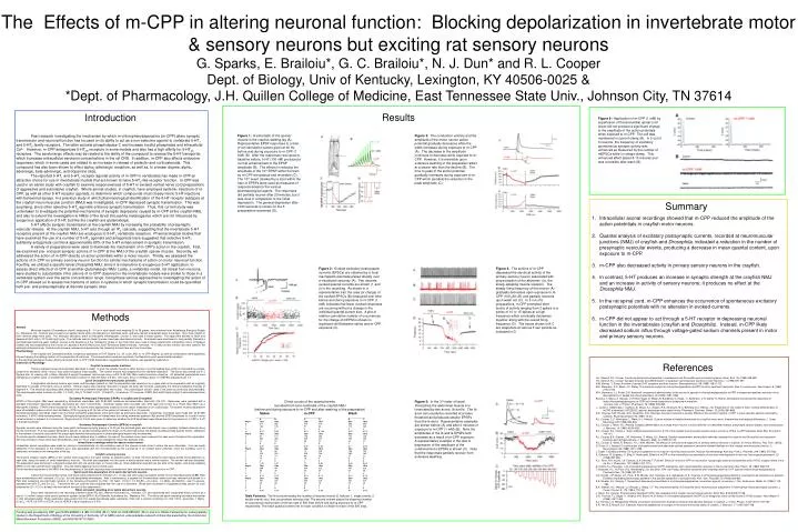
The Effects of m-CPP in altering neuronal function: Blocking depolarization in invertebrate motor & sensory neurons but exciting rat sensory neurons G. Sparks, E. Brailoiu*, G. C. Brailoiu*, N. J. Dun* and R. L. Cooper Dept. of Biology, Univ of Kentucky, Lexington, KY 40506-0025 & *Dept. of Pharmacology, J.H. Quillen College of Medicine, East Tennessee State Univ., Johnson City, TN 37614 Introduction Results Figure 6: Application of m-CPP (1 mM) by superfusion of thoracolumbar spinal cord slices did not produce a significant change in the amplitude of the action potentials when exposed to m-CPP. The cell was maintained in current-clamp (A). In 5 out of 6 neurons, the frequency of excitatory spontaneous synaptic activity was enhanced as measured by the number of mEPSCs while in voltage-clamp. This enhanced effect lasted 8-10 minutes and was reversible after wash (B). Figure 1: A schematic of the opener muscle in the crayfish walking leg (A). Representative EPSP responses to a train of ten stimulation pulses given at 40 Hz before and during exposure to m-CPP (1 mM) (B). After the responses recovered to baseline values, 5-HT (100 nM) produced a normal enhancement of the EPSP amplitude (B). The effects in reducing the amplitude of the 10th EPSP within the train by m-CPP are gradual and reversible (C). The 10th event (marked by a star) within the train of EPSPs were used as measures of responsiveness to the various pharmacological agents. The responses did partially recover after 20 minutes, but it was slow in comparison to the initial depression. The general depression after 1000 seconds is shown for the 5 preparations examined (D). Figure 3: The conduction velocity and the amplitude of the motor neuron action potential gradually decreases while the width increases during exposure to m-CPP (A). The decrease in the amplitude continues to decrease while exposed to m-CPP. However, it is reversible upon extensive washing of the preparation albeit at a slower rate than the decline (B). The time to peak of the action potential gradually increases during exposure to m-CPP which parallels the reduction in the peak amplitude (C). Past research investigating the mechanism by which m-chlorophenylpiperazine (m-CPP) alters synaptic transmission and neuronal function has focused on its ability to act as a non-selective agonist to vertebrate 5-HT1 and 5-HT2-family receptors. The latter activate phospholipase C and increase inositol phosphates and intracellular Ca2+. However, m-CPP antagonizes 5-HT2B receptors in some models and also has a high affinity for 5-HT2C receptors. The serotonergic effects may be related to the ability of the compound to reverse the 5-HT transporter, which increases extracellular serotonin concentrations in the rat CNS. In addition, m-CPP also affects endocrine responses, which in some cases are related to an increase in release of prolactin and corticosteroids. This compound has also been shown to effect alpha2-adrenergic receptors, as well as, to a lesser degree, alpha1-adrenergic, beta-adrenergic, and dopamine sites. The reported 5-HT1 and 5-HT2 receptor agonist activity of m-CPP in vertebrates has made m-CPP an attractive choice for use in invertebrate models that are known to have 5-HT2-like receptor function. m-CPP was used in an earlier study with crayfish to examine responsiveness of 5-HT in excised ventral nerve cord preparations of aggressive and submissive crayfish. Whole animal studies, in crayfish, have employed systemic injections of m-CPP, as well as other 5-HT receptor agonists, to determine which compounds most closely mimic 5-HT injections with behavioral assays. In a previous study in which pharmacological identification of the 5-HT receptor subtypes at the crayfish neuromuscular junction (NMJ) was investigated, m-CPP depressed synaptic transmission. This was surprising, since other selective 5-HT2 agonists enhance synaptic transmission. Thus, this current study was undertaken to investigate the potential mechanisms of synaptic depression caused by m-CPP at the crayfish NMJ, and also to extend the investigation to NMJs of the larval Drosophila melanogaster, which are not influenced by exogenous application of 5-HT, but like the crayfish are glutamatergic. 5-HT affects synaptic transmission at the crayfish NMJ by increasing the probability of presynaptic vesicular release. At the crayfish NMJ, 5-HT acts through an IP3 cascade, suggesting that the invertebrate 5-HT receptors present at the crayfish NMJ are analogous to 5-HT2 vertebrate receptors. Pharmacological studies that have examined the use of a number of 5-HT2 agonists and antagonists have suggested that selective 5-HT2 subfamily antagonists can block approximately 80% of the 5-HT enhancement in synaptic transmission. A variety of preparations were used to illuminate the mechanism of m-CPP’s action in the crayfish. First, we examined pre- and post-synaptic actions of m-CPP at the NMJ of the crayfish opener muscle. Secondly, we addressed the action of m-CPP directly on action potentials within a motor neuron. Thirdly, we assessed the actions of m-CPP on primary sensory neuron function for similar mechanisms of action on motor neuronal function. Fourthly, we utilized a specific larval Drosophila NMJ, since it is insensitive to exogenous 5-HT application, to assess direct effects of m-CPP at another glutamatergic NMJ. Lastly, a vertebrate model, rat dorsal horn neurons, were studied to substantiate if the actions of m-CPP observed in the invertebrate models were similar to those in a vertebrate system over the same concentration range. Using these various approaches to investigating the action of m-CPP allowed us to assess mechanisms of action in systems in which synaptic transmission could be quantified both pre- and postsynaptically at discrete synaptic sites. Summary • Intracellular axonal recordings showed that m-CPP reduced the amplitude of the action potetntials in crayfish motor neurons. • Quantal analysis of excitatory postsynaptic currents, recorded at neuromuscular junctions (NMJ) of crayfish and Drosophila, indicated a reduction in the number of presynaptic vesicular events, producing a decrease in mean quantal content, upon exposure to m-CPP. • m-CPP also decreased activity in primary sensory neurons in the crayfish. • 4. In contrast, 5-HT produces an increase in synaptic strength at the crayfish NMJ and an increase in activity of sensory neurons; it produces no effect at the Drosophila NMJ. • 5. In the rat spinal cord, m-CPP enhances the occurrence of spontaneous excitatory postsynaptic potentials with no alteration in evoked currents. • 6. m-CPP did not appear to act through a 5-HT receptor in depressing neuronal function in the invertebrates (crayfish and Drosophila). Instead, m-CPP likely decreased sodium influx through voltage-gated sodium channels present in motor and primary sensory neurons. Figure 2: Evoked excitatory postsynaptic currents (EPSCs) are obtained by a focal macropatch electrode placed directly over a visualized varicosity (A). Two discrete evoked quantal currents are shown (1 and 2) in the recording. As shown in a representative trail, the area (or charge) of the evoked EPSCs (B) measured over time before and during exposure to m-CPP (1 mM) indicates that fewer evoked responses are occurring without a change in the individual quantal current size. A plot of relative cumulative number of occurrences for the charge of mEPSCs shows no significant shift between saline and m-CPP exposure (C). Figure 4: The actions of m-CPP depressed the electrical activity of the primary sensory neuron associated with proprioception of the abdomen (i.e. the slowly-adapting muscle receptor). The steady firing frequency of the neuron (A) gradually decreases upon exposure to m-CPP (100 mM) (B) and partially recovers upon wash out (C). In 5 out of 5 preparations, m-CPP prompted short bursts of activity ranging from 2 spikes to a series of 10 or 15 spikes at a high frequency which eventually decreases together along with the overall firing frequency (D). The traces shown in A-C are snapshots at various 5 sec periods as indicated in D. Methods Animals Mid-sized crayfish (Procambarus clarkii), measuring 8 - 10 cm in body length and weighing 20 to 36 grams, were obtained from Atchafalaya Biological Supply Co. (Raceland, LA). Animals were housed in an aquatic facility within the laboratory in individual tanks, and were fed fish food pellets every three days. Only male crayfish in their intermolt stage were used. The ‘wild-type’ laboratory strain of Drosophila melanogaster, Canton S, was used in these studies. The eggs were allowed to hatch and develop at 25oC with a 12:12 dark-light cycle. The methods used to stage fly larvae have been described previously. All animals were maintained in vials partially filled with a cornmeal-agar-dextrose-yeast medium. Larvae at the beginning of the “wandering” phase of the third instar were used in these experiments. A breeding colony of Sprague Dawley rats was established at the Division of Laboratory Animal Resources, East Tennessee State University. Immature, 10-15-day-old rats of either sex were used for the electrophysiological study. Animal protocols were reviewed and approved by the University Animal Care and Use Committee. Pharmacology In the crayfish and Drosophila studies, exogenous application of 5-HT (Sigma Co., St. Louis, MO) or m-CPP (Sigma), as well as combinations were applied by fully exchanging the bathing medium of the preparation three times. The concentrations used are reported in the Results for each experimental paradigm. In the electrophysiological studies utilizing rat spinal cord, m-CPP (1mM) dissolved in oxygenated Krebs solution, was applied by superfusion. Dissection & Physiology Crayfish neuromuscular junctions These procedures have previously been described in detail. In brief, the opener muscle in either the first or second walking legs, which is innervated by a single, purely-tonic excitatory motor neuron, was used throughout these studies. The opener muscle was prepared by the standard dissection. The tissue was pinned out in a Slygard dish for viewing with a Nikon Optiphot‑2 upright fluorescent microscope using a 40X (0.55 NA) Nikon water-immersion objective. All dissected preparations were maintained in crayfish saline, a modified Van Harreveld's solution (in mM: 205 NaCl; 5.3 KCl; 13.5 CaCl2 2H2O; 2.45 MgCl26H2O; 0.5 HEPES) adjusted to pH 7.4. Larval Drosophila neuromuscular junctions A longitudinal mid-dorsal incision was made, and the edges pinned, so that the preparation was spread out on a glass slide in the preparation dish as originally described for studies of the leech nervous system. Internal organs were carefully removed to expose the body wall muscles, particularly the ventral longitudinal muscles of segment 4. The electrical recordings were obtained from the prominent longitudinal m6 muscle. The physiological solution used is the same as previously described [80]. The physiological saline contains (in mM): 1.0 CaCl2 .2H2O, 70 NaCl, 5 KCl, 10 NaHCO3, 5 trehalose, 115 sucrose, 5 BES (N,N-bis[2-Hydroxyethyl]-2-aminoethanesulfonic acid). Excitatory Postsynaptic Potentials (EPSPs) in crayfish and Drosophila EPSPs at the crayfish NMJ were recorded by intracellular electrodes, with 30‑60 MΩ resistance microelectrodes, filled with 3 M KCl. Responses were recorded with a standard intracellular electrode amplifier (AxoClamp 2A, Axon Instruments). Electrical signals were recorded onto VHS tape and on‑line to a Power Mac 9500 via a MacLab/4s interface. EPSPs were recorded at 10 kHz. All events were appropriately scaled to known values measured on an oscilloscope. The opener muscle preparations were stimulated to induce a short-term facilitation (STF) by giving a 40 Hz train of ten pulses at intervals of 5 or 10 seconds. Electrophysiological recordings made from the larval Drosophila preparations were performed as previously described. Intracellular recordings were made with 30‑60 MΩ resistance, 3 M KCl-filled microelectrodes. Electrophysiological parameters of interest were the resting membrane potential (Rp) and the EPSP amplitudes for Is and Ib motor nerve terminals in segment 4 of muscle m6. Single stimuli at 0.5 Hz were given to determine whether the EJP amplitudes were altered due to the presence of m-CPP or 5-HT in the bathing media. Excitatory Postsynaptic Currents (EPSCs) in crayfish Synaptic currents were obtained using the loose patch technique by lightly placing a 10-20 μm fire polished glass electrode directly over a spatially isolated varicosity along the nerve terminal. The macropatch electrode is specific for current recording within the region of the electrode lumen. By directly counting evoked quantal events, alterations in the number of vesicles fusing within the presynaptic terminal during exposure to pharmacological agents may be observed. To monitor quanta released over time, direct counts were obtained and, in addition, the area of the evoked current was measured for each event throughout the experiment. The tonic excitatory motor nerve was stimulated at a rate of 1 Hz in order not to facilitate the responses between trials. Intracellular recordings within the opener motor neuron Intracellular axonal recordings were made by placing a microelectrode into the excitatory axon of the opener muscle close to where the axon bifurcates. One can easily determine if the excitatory or the inhibitory axon was penetrated with the microelectrode by the occurrence of an evoked action potential, since the excitatory axon is selectively stimulated in the meropodite of the leg. Crayfish sensory neurons The muscle receptor organs (MRO) of the crayfish were exposed in the same manner as detailed earlier. In brief, the shell along the lower lateral border of the abdomen on each side, along the series of small indentations, was cut. The shell was separated into two parts--a dorsal section and a ventral section. The ventral half was discarded. The preparation was anchored to a Sylgard-coated dish with the ventral view, or muscle-side-up. Each abdominal segment has two sets of the rapidly- and slowly-adapting MROs on the right and left hemi-segments. Only the slowly-adapting neurons were used. For the sensory responses of the MRO, the firing frequency of the static response was monitored over time before and during exposure to m-CPP. Spinal cord slice preparation of rat spinal dorsal horn neurons Transverse thoracolumbar spinal cord slices were prepared from immature (5 – 10 day-old) rats using a procedure similar to that described earlier [2,88]. Rats were anaesthetized with urethane (1.2 g kg-1,ip) and decapitated immediately. After a laminectomy, a segment of thoracolombar spinal cord was removed and placed in a Petri dish containing ice-cold Krebs solution of the following composition (in mM): 127 NaCl, 1.9 KCl, 1.2 KH2PO4, 2.4 CaCl2, 1.3 MgCl2,26 NaHCO3, and 10 glucose, saturated with 95%O2 and 5% CO2. Transverse 400 m sections were prepared with the use of a vibrotome. Slices were incubated in oxygenated Krebs solution at room temperature (211C) for at least one hour before the start of the experiments. Whole cell patch recording of rat spinal dorsal horn neurons Slices were transferred to the recording chamber (model RC-22C, Warner Instrument Inc., Hamden, CT) and superfused with oxygenated Krebs solution at a rate of 1-2 ml/min using a valve control perfusion system (model BPS-4; ALA Scientific Instruments Inc., Westbury, NY). The whole cell patch recording technique was similar to that described earlier. Patch electrodes were pulled from thin-walled borosilicate glass capillaries, filled with a solutioncontaining (in mM) 130 K- gluconate, 1 MgCl2, 2 CaCl2, 4 ATP, 0.3 GTP,10 EGTA, and 10 HEPES, had a resistance of 3-5 M. References H.L. Atwood, R.L. Cooper, Functional and structural parallels in crustaceans and Drosophila neuromuscular systems, Amer. Zool. 35 (1995) 556-565. H.L. Atwood, R.L. Cooper, Synaptic diversity and differentiation: Crustacean neuromuscular junctions, Invert. Neurosci. 1 (1996) 291-307. N.M. Barnes, T. Sharp, A review of central 5-HT receptors and their function, Neuropharmacol. 58 (1999) 1083-1152. M.H. Baumann, D.C. Mash, J.K. Staley, The serotonin agonist m-chlorophenylpiperazine (mCPP) binds to serotonin transporter sites in human brain, Neuroreport. 6 (1995) 2150-2152. M.H. Baumann, J.J. Rutter, S.B. Auerbach, Intravenous administration of the serotonin agonist m-chlorophenylpiperazine (mCPP) increases extracellular serotonin in the diencephalons of awake rats, Neuropharmacol. 32 (1993) 1381-1386. Broocks, T. Meyer, A. George, U. Hillmer‑Vogel, D. Meyer, B. Bandelow, G. Hajak, U. Bartmann, C.H. Gleiter, E. Ruther, Decreased neuroendocrine responses to meta‑chlorophenylpiperazine (m‑CPP) but normal responses to ipsapirone in marathon runners, Can. J. Physiol. Pharmacol. 76 (1998) 278‑283. L. Castro, I. Maldonado, I. Campos, B. Varjao, A.L. Angelo, R.A. Athanazio, M.C. Barbetta, A.C. Ramos, J.B. Fregoneze, E. De Castro e Silva, Central administration of mCPP, a serotonin 5-HT(2B/2C) agonist, decreases water intake in rats, Pharmacol. Biochem. Behav. 72 (2002) 891-898. D.S. Charney, S.W. Woods, W.K. Goodman, G.R. Heninger, Serotonin function in anxiety. Effects of the serotonin agonist, mCPP, in panic disorder patients and healthy subjects, Psychopharmacol. 92 (1987) 14-24. R.L. Cooper, C.C. Harrington, L. Marin, H.L. Atwood, Quantal release at visualized terminals of a crayfish motor axon: Intraterminal and regional differences, J. Comp. Neurology 375 (1996) 583-600. R.L. Cooper, L. Marin, H.L. Atwood, Synaptic differentiation of a single motor neuron: conjoint definition of transmitter release, presynaptic calcium signals, and ultrastructure, J. Neurosci. 15 (1995) 4209-4222. R.L. Cooper, R.J. Chase, J. Tabor, Altered responsiveness to 5-HT at the crayfish neuromuscular junction due to chronic p-CPA & m-CPP treatment, Brain Res. 916 (2001) 143‑151. R.L. Cooper, B.A. Stewart, J.M. Wojtowicz, S. Wang, H.L. Atwood, Quantal measurement and analysis methods compared for crayfish and Drosophila neuromuscular junctions and rat hippocampus. J. Neurosci. Meth. 61 (1995) 67-78. R.L. Cooper, E. Ward, R. Braxton, H. Li, W.M. Warren, The effects of serotonin and ecdysone on primary sensory neurons in crayfish, (In Press, Microsc. Res. Tech. (2003). D. Dixon, H.L. Atwood, Conjoint action of phosphoinositol and adenylate cyclase systems in serotonin-induced facilitation at the crayfish neuromuscular junction, J. Neurophysiol. 62 (1989) 1251-1259. J. Dudel, Facilitatory effects of 5-hydroxy-tryptamine on the crayfish neuromuscular junction, Naunyn-Schmiedebergs Arch.exp. Path.u. Pharmak. 249 (1965) 515-528. E. Eriksson, G. Engberg, O. Bing, H. Nissbrandt, Effects of mCPP on the extracellular concentrations of serotonin and dopamine in rat brain, Neuropsychopharmacol. 20 (1999) 287‑296. K.C. Fone, R.H. Austin, I.A. Topham, G.A. Kennett, T. Punhani, Effect of chronic m‑CPP on locomotion, hypophagia, plasma corticosterone and 5‑HT2C receptor levels in the rat, Br. J. Pharmacol. 123 (1998) 1707‑1715. A. Hamik, S.J. Peroutka, 1-(m-Chlorophenyl)piperazine (mCPP) Interactions with neurotransmitter receptors in the human brain, Biol. Psych. 25 (1989) 569-575. T. Klaassen, K.L. Ho Pian, H.G. Westenberg, J.A. den Boer, H.M. van Praag, Serotonin syndrome after challenge with the 5‑HT agonist meta‑chlorophenylpiperazine, Psychiatry Res. 79 (1998) 207‑212. J.H. Krystal, J.P. Seibyl, L.H. Price, S.W. Woods, G.R. Heninger, G.K. Aghajanian, D.S. Charney, m-Chlorophenylpiperazine effects in neuroleptic-free schizophrenic patients. Evidence implicating serotonergic systems in the positive symptoms of schizophrenia, Arch. Gen. Psychiatr. 50 (1993) 624-635. E.A. Mueller, D.L. Murphy, T. Sunderland, Neuroendocrine effects of m-chlorophenylpiperazine, a serotonin agonist, in humans, J. Clin. Endocrinol. Metab. 61 (1985) 1179-1184. B.A. Stewart, H.L. Atwood, J.J. Renger, J. Wang, C.F. Wu, Improved stability of Drosophila larval neuromuscular preparation in haemolymph-like physiological solutions, J. Comp. Physiol. A. 175 (1994) 179-191. J. Tabor, R.L. Cooper, Physiologically identified 5-HT2 -like receptors at the crayfish neuromuscular junction, Brain Res. 932 (2002) 91-98. D.R. Thomas, T.L. Gager, V. Holland, A.M. Brown, M.D. Wood, m-Chlorophenylpiperazine (mCPP) is an antagonist at the cloned human 5-HT2B receptor, NeuroReport 7 (1996) 1457-1460. A.J. Tierney, L.A. Mangiamele, Effects of serotonin and serotonin analogs on posture and agonistic behavior in crayfish, J. Comp. Physiol. A 187 (2001) 757-767. S.R. Yeh, B.E. Musolf, D.H. Edwards, Neuronal adaptations to changes in the social dominance status of crayfish, J. Neurosci. 17 (1997) 697-708. Direct counts of the quantal events recorded from tonic terminals of the crayfish NMJ (before and during exposure to m-CPP and after washing of the preparation) Saline m-CPP Wash _________________ ________________ _______________ A B C D E F Prep 1 Obs Obs Obs Obs Obs Obs 0 317 353 415 416 380 394 1 164 145 91 84 115 104 2 9 2 3 0 5 2 m 0.37 0.29 0.19 0.168 0.25 0.22 Prep 2 Obs Obs Obs Obs Obs Obs 0 87 113 145 205 176 120 1 392 368 329 273 298 352 2 21 19 25 22 24 27 3 0 0 1 0 2 1 m 0.87 0.81 0.76 0.63 0.7 0.82 Prep 3 Obs Obs Obs Obs Obs Obs 0 75 86 74 228 188 199 1 329 333 320 238 285 280 2 76 73 89 28 27 21 3 14 8 16 4 0 0 4 4 0 1 0 0 0 m 1.09 1.01 1.1 0.62 0.68 0.64 Prep 4 Obs Obs Obs Obs Obs Obs 0 400 401 434 422 435 426 1 88 70 63 64 64 68 2 2 0 3 2 1 6 m 0.19 0.15 0.14 0.12 0.13 0.16 Prep 5 Obs Obs Obs Obs Obs Obs 0 457 431 466 470 452 474 1 43 68 26 18 45 25 2 0 1 3 3 3 1 3 0 0 0 1 0 0 m 0.09 0.14 0.07 0.06 0.1 0.05 Table Footnote: The first column states the number of discrete events (0, failures; 1, single events; 2 double events; etc.) that occurred per stimulus trial. The second column states the observed number of occurrences during each of the two sets of 500 trials before and during exposure to m-CPP respectively. The mean quantal content (m) for each condition is shown for each of the 500 trials. Figure 5: In the 3rd instar of larval Drosophila, the abdominal muscle 6 is innervated by two axons, Ib and Is. The Ib axon can usually be recruited at a lower threshold and produces smaller EPSPs than the Is axon. Representative examples are shown before (A) and after 2 minutes of exposure to m-CPP (1 mM) (B). Note the amplitudes of the Ib and Is EPSPs both decrease as a result of m-CPP exposure. A representative example in the rate of depression of the amplitude of the combined Ib+Is EPSPs is shown (C). Note that the responses partially recover from extensive washing. Funding was provided by NSF grants IBN-9808631 & IBN-0131459 (RLC), NSF-ILI-DUE 9850907 (RLC) and a G. Ribble Fellowship for undergraduate studies in the Department of Biology at the University of Kentucky (JT & GMS) and an undergraduate research scholarship awarded by the Arnold and Mabel Beckman Foundation (GMS), and NIH NS18710 (NJD).