Download
1 / 45
510 likes | 815 Views
CHAPTER 9. The Cytoskeleton and Cell Motility. Introduction. The cytoskeleton is a network of filamentous structures: microtubulues , microfilaments , and intermediate filaments. Properties of cytoskeletal components. 9.1 Overview of the Major Functions of the Cytoskeleton (1).

E N D
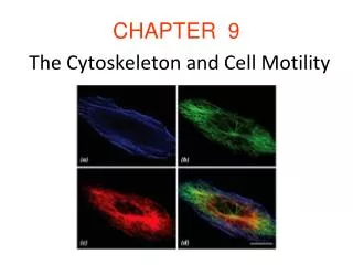
CHAPTER 9 The Cytoskeleton and Cell Motility
Introduction • The cytoskeleton is a network of filamentous structures: microtubulues, microfilaments, and intermediate filaments.
9.1 Overview of the Major Functions of the Cytoskeleton (1) • The cytoskeleton has many roles: • Serves as a scaffold providing structural support and maintaining cell shape. • Serves as an internal framework to organize organelles within the cell. • Directs cellular locomotion and the movement of materials within the cell.
Overview of the Major Functions of the Cytoskeleton (2) • Provides anchoring site for mRNA. • Serves as a signal transducer. • An essential component of the cell’s division machinery.
9.2 Study of the Cytoskeleton (1) • The Use of Live-Cell Fluorescence Imaging • Can be used to locate fluorescently-labeled target proteins. • Molecular processes can be observed (live-cell imaging). • Used to reveal the location of a protein present in very low concentrations.
Study of the Cytoskeleton (2) • The Use of In Vitro Single-Molecule Assays • They make possible to detect the activity of an individual protein molecule in real time. • Can be supplement with atomic force microscopy to measure the mechanical properties of cytoskeletal elements.
Using video microscopy to follow activities of molecular motors
9.3 Microtubules (1) • Structure and Composition • Microtubules are hollow, cylindrical structures. • The microtubule is a set of globular proteins arranged in longitudinal rows called protofilaments. • Microtubules contain 13 protofilaments. • Each protofilament is assembled from dimers of α- and ß-tubulin subunits assembled into tubules with plus and minus ends.
Microtubules (2) • Microtubule-Associated Proteins (MAPs) • MAPs comprise a heterogeneous group of proteins. • MAPs attach to the surface of microtubules to increase their stability and promote their assembly. • MAPs are regulated by phosphorylation of specific amino acid residues.
Microtubules (2) • Microtubules as Structural Supports and Organizers • The distribution of microtubules determines the shape of the cell. • Microtubules maintain the internal organization of cells.
Microtubules (3) • Microtubules as Structural Supports and Organizers • Microtubules function in axonal transport. • Microtubules play a role in axonal growth during embryogenesis.
Microtubules (4) • Microtubules as Structural Supports and Organizers • In plant cells, microtubules help maintain cell shape by influencing formation of the cell wall.
Microtubules (5) • Microtubules as Agents of Intracellular Motility • They facilitate movement of vesicles between compartments. • Axonal transport: • Movement of neurotransmitters across the cell. • Movement away from the cell body (anterograde) and toward the cell body (retrograde). • Mediate tracks for a variety of motor proteins.
Microtubules (6) • Motor Proteins that Traverse the Microtubular Cytoskeleton • Molecular motors convert energy from ATP into mechanical energy. • Molecular motors move unidirectionally along their cytoskeletal track in a stepwise manner. • Three categories of molecular motors: • Kinesin and dynein move along microtubule tracks. • Myosin moves along microfilament tracks.
Microtubules (7) • Kinesins • Kinesin—member of a superfamily called KLPs (kinesin-like proteins). • A kinesin is a tetramer of two identical heavy chains and two identical light chains. • Each kinesin includes a pair of globular heads (motor domain), connected to a rod-like stalk. • Kinesin is a plus end-directed microtubular motor based on its movement.
Microtubules (8) • Kinesins (continued) • They move along a single protofilament of a microtubule at a velocity proportional to the ATP concentration. • Movement is processive, motor protein moves along an individual microtubule for a long distance without falling off. • KLPs move cargo toward the cell’s plasma membrane.
Microtubules (9) • Cytoplasmic Dynein • Dynein – responsible for the movement of cilia and flagella. • Cytoplasmic dynein – Huge protein with a globular, force-generating head. • It is a minus end-directed microtubular motor. • Requires an adaptor (dynactin) to interact with membrane-bounded cargo.
Microtubules (10) • Microtubule-Organizing Centers (MTOCs) • MTOCs – specialized structures for the nucleation of microtubules. • Centrosome – structures responsible for initiating microtubules in animal cells. • It contains twobarrel-shaped centrioles surrounded by pericentriolar material (PCM). • Centrioles are usually found in pairs.
Microtubules (11) • Centrosomes (continued) • Responsible for initiation and organization of the microtubular cystoskeleton. • Microtubules terminate in the PCM.
Microtubules (12) • Basal Bodies and Other MTOCs • Basal body – structure where outer microtubules in a cilia and flagella are generated. • Plant cells lack MTOCs and their microtubules are organized around the surface of the nucleus.
Microtubules (13) • Microtubule Nucleation • MTOCs control the number of microtubules, their polarity, the number of protofilaments, and the time and location of their assembly. • The protein -tubulin is found in all MTOCs and is critical for microtubule nucleation.
Microtubules (14) • The Dynamic Properties of Microtubules • There are four distinct arrays of microtubules in a dividing plant cell: • Widely distributed throughout the cortex. • Making a single transverse band. • In the form of a mitotic spindle. • As a phargmoplast assisting in the formation of the cell wall of daughter cells.
Microtubules (15) • The Dynamic Properties of Microtubules • Newly formed microtubules branch at an angle of pre-existing microtubules. • The changes in spatial organization of microtubules are a combination of two mechanisms: • Rearrangement of existing microtubules. • Disassembly of existing microtubules and reassembly of new one in different locations.
Microtubules (16) • The Underlying Basis of Microtubule Dynamics • Insight into factors that influence microtubule assembly and disassembly came from studies in vitro. • GTP is required for microtubule assembly. • Hydrolysis of GTP leads to a replacement of bound GDP by new GTP to “recharge” the tubulin dimer.


