Download
1 / 3
30 likes | 153 Views
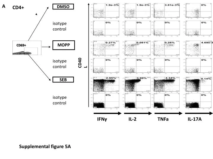
IFN γ. IL-2. TNFa. IL-17A. A. CD4+. DMSO. isotype control. MOPP. CD69+. CD40L. isotype control. SEB. isotype control. Supplemental figure 5A. IFN γ. IL-2. TNFa. IL-17A. B. CD8+. DMSO. isotype control. MOPP. isotype control. CD69. SEB. isotype control.

E N D
IFNγ IL-2 TNFa IL-17A A CD4+ DMSO isotype control MOPP CD69+ CD40L isotype control SEB isotype control Supplemental figure 5A
IFNγ IL-2 TNFa IL-17A B CD8+ DMSO isotype control MOPP isotype control CD69 SEB isotype control Supplemental figure 5B
Supplemental Figure S5. Isotype controls for CD4+ (A) and CD8+ (B) T-cell responses Gates were set on CD3+ lymphocytes, doublets and dead cells were discriminated. CD4+ (A) and CD8+ (B) T-cells were subdivided. CD69 was used as an early activation marker and CD40L - as a marker for T- helper cells which have been recently activated. Subsequent multiparameter flow cytometric analysis was performed according to the expression of IFNγ, IL-2, TNFa and IL-17A. SEB was used as positive control and DMSO as negative control. CD69+ gating strategy was chosen as a representative sample after SEB stimulation. Isotype-matched control antibodies were used to distinguish non-specific (“background”) stainings.