Download
1 / 1

E N D
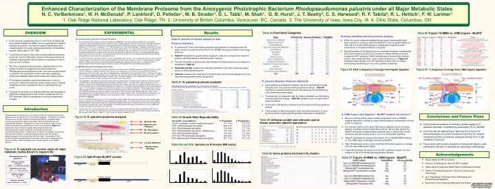
Enhanced Characterization of the Membrane Proteome from the Anoxygenic Phototrophic Bacterium Rhodopseudomonas palustris under all Major Metabolic StatesN. C. VerBerkmoes1, W. H. McDonald1, P. Lankford1, D. Pelletier1, M. B. Strader1, D. L. Tabb1, M. Shah1, G. B. Hurst1, J. T. Beatty2, C. S. Harwood3, R. F. Tabita4, R. L. Hettich1, F. W. Larimer11. Oak Ridge National Laboratory, Oak Ridge, TN2. University of British Columbia, Vancouver, BC, Canada 3. The University of Iowa, Iowa City, IA4. Ohio State, Columbus, OH EXPERIMENTAL OVERVIEW Results Table #4 Functional Categories Table #8 Trypsin 1D-MMS vs. CNBr/trypsin - MudPIT Problems identified with the proteome analysis. • While the current proteome analysis shows quality reproducibility and large numbers of unique proteins identified from various growth states it is clear that the current methodology is inadequate at analyzing some components of integral membrane complexes. • Detailed analysis of the results from the membrane fractions indicated that while some proteins known to be involved in integral membrane protein complexes (ATP synthase, NADH hydrogenase, photosynthetic reaction center), were easily identified, other components were not. Figure #4 highlights total coverage over ATP synthase complex with the trypsin methodology illustrating just one of the problem complexes. Initial R. palustris Proteome analysis to date: Proteome Statistics: • R. palustris WT and LhaA mutant proteome were analyzed in duplicate under all major modes of growth by automate 1D-LC-MS/MS employing multiple mass range scanning. • Table #1 highlights the growth states analyzed to data and compares the total # of proteins identified based at different levels of filtering. • The reproducibility of each growth state between the duplicate proteome analysis is illustrated in Table #2. • Table #3a and #3b compare the observed distribution of protein molecular weight and pI vs the predicted genome. • Table #4 compares the observed # of proteins from the functional categories vs. the total # proteins predicted from the genome. Cell Growth and Production of Protein Fractions: • All major growth states analyzed to date are highlighted in Figure #2. R. palustris CGA010 wild-type strain was grown anaerobically (photoheterotrophic growth) in light to mid-log or stationary phase, and the WT stain and a LhaA (light harvesting apparatus assembly protein) mutant were grown aerobically (chemoheterotrophic growth) with shaking, in defined mineral medium at 30°C(Kim, M.-K. & Harwood, C. S.; FEMS Microbiol. Lett. 1991) to mid-log phase. Ammonium sulfate and succinate were provided as a nitrogen and carbon source for all of these growth states. For nitrogen fixation conditions the wild-type strain was grown anaerobically as a above with N2 gas continually bubbling through the media as the sole source of nitrogen. For photoautotrophic growth the wild-type strain was grown anaerobically as above with CO2 gas continually bubbling through the media as the sole source of carbon. The benzoate growth state was the same as the anaerobic state above except benzoate was substituted for succinate as the sole carbon source. • Cells were harvested, washed twice with Tris buffer, and disrupted with sonication. Four crude protein fractions were created by ultracentrifugation (100,000g for 1 hour creates membrane and crude fraction and then for 24 hours creates pellet and cleared fraction). Protein fractions were denatured, reduced and digested with sequencing grade trypsin. In separate analysis photoautotrophic and chemoheterotrophic membrane fractions were digested with a dual CNBr/trypsin digestion. LC-MS/MS Analysis and Database searching: • All tryptic digestions of all growth states were analyzed via one-dimensional LC-MS/MS experiments performed with an Ultimate HPLC (LC Packings, a division of Dionex, San Francisco, CA) coupled to an LCQ-DECA or LCQ-DECA XP ion trap mass spectrometer (Thermo Finnigan, San Jose, CA) equipped with an electrospray source operated at 4.5kV. Injections were made with a Famos (LC Packings) autosampler onto a 50μl loop. Flow rate was ~4μL/min with a 240-min gradient for each LC-MS/MS run. A VYDAC 218MS5.325 (Grace-Vydac, Hesperia, CA) C18 column (300μm id x 15cm, 300Å with 5μm particles) was directly connected to the Finnigan electrospray source with 100μm id fused silica. • The CNBr/trypsin dual digest of the photoautotrophic and chemoheterotrophic membranes were analyzed via two-dimensional LC-MS/MS with a split-phase MudPIT column described in McDonald, W.H.; et al. IJMS, 2002. Approximately 3cm of SCX material (Luna SCX 5μm 100A Phenomenex) was first packed into a 100μm fused silica via a pressure cell followed by 3cm of C-18 RP material (Aqua C-18 5μm 200A Phenomenex, Torrance, CA). The sample was then loaded off-line onto the dual phase column. For this study two separate concentrations were tested for each sample (concentrated ~500μg starting material, dilute ~50μg starting material). The RP-SCX column was then positioned on the instrument behind a ~12cm c18 RP column (Jupiter 5um 300A Phenomenex also packed by pressure cell into Pico Frit tip (75μm with 15μm tip New Objective, Woburn, MA) positioned directly in the nanospray source on a LCQ-DECA (nanospray voltage 2.2kV). The samples were analyzed via a 24-hour MudPIT analysis detailed in Washburn; et al. Nature Biotech. 2001. The column configuration for this experiment is illustrated in Figure #3. • For all LC/MS/MS data acquisition, the LCQ was operated in the data dependent mode with dynamic exclusion enabled, where the top four peaks in every full MS scan were subjected to MS/MS analysis. To increase dynamic range in the 1D-LC-MS/MS analysis separate injections were made with a total of 8 overlapping segmented m/z ranges scanned (referred to as gas phase fractionation or multiple mass range scanning MMS). All samples digested with trypsin and analyzed via 1D-LC-MS/MS with multiple mass range scanning were run in duplicate. • The resultant ~500 .raw files from all proteome analyses were searched with SEQUEST against all predicted ORFs from R. palustris. The raw output files were filtered and sorted with DTASelect and growth states and sample to sample reproducibility were compared with Contrast (Tabb, D.L.; et al, Journal of Proteome Research, 2003). • In the recently created Genomes to Life Center for Molecular and Cellular Systems at ORNL, the core goal will be to build a research program for the high-throughput identification and characterization of protein complexes primarily from bacterial species (See poster # ThPT 388). • To achieve this goal for any new microbial species a baseline proteome must be obtained to ensure that target proteins are selected in appropriate manner based on expression under a given growth condition. • We have completed initial proteome characterization of the purple nonsulfur anoxygenic phototrophic bacteriumRhodopseudomonas palustris under a all major growth modes to ascertain the expression profiles and major qualitative differences between these growth states and mutant strains. • This analysis revealed a significant weakness in current protocols to effectively analysis certain components of integral membrane protein complexes known to expressed at high levels. • The goal of this study is to evaluate different methodologies to effectively analyze these known protein complexes such as ATP synthase, NADH hydrogenase, and the photosynthetic complex. Figure #5 % Sequence Coverage from CNBr/trypsin digestion Figure #4 Total % Sequence Coverage from trypsin digestion R. palustris Baseline Proteome Highlights • Initial qualitative comparision between Aerobic and Anaerobic growth indicated over 120 proteins showing significant change. Table #5 highlights a unknown protein and an interesting operon identified only under anaerobic growth states. • The Anaerobic vs. Anaerobic with N2 fixation indicated over 50 proteins showing significant change. Table #6 highlights some nitrogen fixation specific proteins. • Autotrophic vs Anaerobic indicated over 80 proteins showing significant change. • Initial analysis of Benzoate growth state indicates expression of most known proteins in benzoate and aromatic hydrocarbon metabolism. Table #1 R. palustris proteome analyzed INTRODUCTION Introduction Is CNBr/trypsin dual digestion - MudPIT analysis the solution? • We are currently testing various sample preperation and LC-MS/MS analysis methods in attempt to clearly identify missing components of known protein complexes. • Our first test method is the dual CNBr/trypsin digestion due to its known ability to solubilize and cut membrane proteins. We are also testing the MudPIT technique to analyze these samples since it offers better overall sensitivity and less surface area to lose very hydrophobic peptides. • Table #7 illustrates the results of the trypsin 1D-LC-MS/MS MMS analysis vs. the CNBr/trypsin dual digest followed by MudPIT. • Table #8 highlights some proteins identified with better sequence coverage from the dual CNBr/trypsin digest. • Figure #5 illustrates the results from the ATP synthase complex from the analysis of the concentrated photoautotrophic sample. Rhodopseudomonas palustris is a purple nonsulfur anoxygenic phototrophic bacterium that is ubiquitous in soil and water samples. R. palustris is of great interest due to its high metabolic diversity and ability to degrade simple aromatic hydrocarbons (lignin monomers). While many bacterium are metabolically versatile, R. palustris is unique in its ability to catalyze more cellular processes than probably any known living organism (Figure #1). Specifically, this microbe is capable ofphotoheterotrophic and photoautotrophic growth, as well as chemoheterotrophic and chemoautotrophic growth. Furthermore, R. palustris is capable of producing hydrogen gas making it a potential biofuel producer and can act as a greenhouse gas sink by converting CO2 into cells. The genome of this microbe had been completed and annotated (Larimer et al, Nature Biotech, 2004) revealing ~4836 potential protein encoding genes in a 5.459Mb genome. Conclusions and Future Plans Figure #2 R. palustris proteome analyzed Table #2 Growth State Reproducibility Table #5 Unknown protein and unknown operon shown anaerobic specific expression. • Initial proteome analysis of all major growth states of R. palustris has been completed by automated 1D-LC-MS/MS. • Currently we are attempting to optimize the protocol for efficient analysis of current membrane fractions for integral membrane protein complexes analyzed directly from crude membrane fractions. • Future work will include completion of all growth states crude membrane fractions in duplicate by optimized methodology. Table #3a and #3b Genome vs Proteome MW and pI Figure #1 R. palustris can survive under all major metabolic modes known to support life Table #6 Some proteins involved in N2 fixation Acknowledgements Figure #3 Split-Phase MudPIT column Table #7 Trypsin 1D-MMS vs. CNBr/trypsin - MudPIT • Grace Vydac for HPLC columns • Dionex-LC Packings for nano 2D HPLC system • Yates Lab at Scripps and David Tabb for DTASelect/Contrast • ORNL-UT Graduate program in Genomic Science and Technology • U.S. Department of Energy Office of Biological and Environmental Research • Genomes to Life Project and Microbial Cell Project