Download
1 / 76
770 likes | 1.19k Views
Lymphatic Filariasis. B.Ganesh Regional Filaria Training & Research Centre National Institute of Communicable Diseases Kozhikode. Lymphatic Filariasis. Infection with 3 closely related Nematodes Wuchereria bancrofti Brugia malayi Brugia timori

E N D
Lymphatic Filariasis B.Ganesh Regional Filaria Training & Research Centre National Institute of Communicable Diseases Kozhikode.
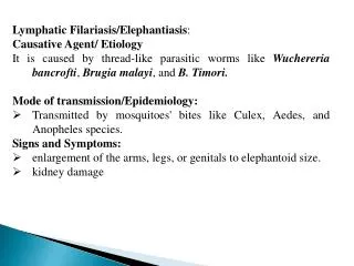
Lymphatic Filariasis Infection with 3 closely related Nematodes • Wuchereria bancrofti • Brugia malayi • Brugia timori * Transmitted by the bite of infected mosquito responsible for considerable sufferings/deformity and disability * All the parasites have similar life cycle in man * Adults seen in Lymphatic vessels * Offsprings seen in peripheral blood during night
Disease Manifestation Disease manifestation range from • None • Acute-Filarial fever • Chronic-Lymphangitis, Lymphadenitis, Elephantiasis of genitals/legs/arms • Tropical Pulmonary Eosinophilia (TPE) • Filarial arthritis • Epididimoorchitis • Chyluria, etc.
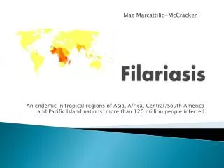
Distribution • Prevalent world wide in the Tropics and Sub-tropical regions of • Africa • Asia • Western Pacific • Parts of Central & South America
Lymphatic Filariasis Endemic Countries & Territories Endemic Countries Global Distribution Map
Global Scenario • Population at risk : 1.2 Billion • No. of countries : > 80 • Mf carriers : 76 Million • Diseased : 44 Million • Hydrocele : 27 Million • Lymphoedema : 16 Million • TPE : 1 Million
National Scenario • Total Population : 110 C • Population at risk : 45.4 C (in 16 States & 5 UT’s) • Total infected : 51.7 M (Wb - 99.4 % and Bm - 0.6 %) • No. of diseased : 22.5 M • Mf carriers : 29.2 M • Hydrocele : 12.9 M
Host Factors • Man – Natural Host • Age – All age (6 months) Max: 20-30 years • Sex – Higher in men • Migration – leading to extension of infection to non-endemic areas • Immunity – may develop after long year of exposure (Basis of immunity-not known)
Social & Environmental Factors • Associated with Urbanization, Poverty, Industrialization, Illiteracy and Poor sanitation. • Climate: is an important factor which influences: • The breeding of mosquito • Longevity (Optimum temperature 20-300C & Humidity 70%) • The development of parasite in the vector • Sanitation, Town planning, Sewage & Drainage.
Mode of Transmission & Incubation Period • Lymphatic Filariasis is transmitted by the bite of Infected mosquito which harbours L3 larva. • L1: 1-3 hours • L2: 3-4 days • L3: 5-6 days • Pre-patent period: (L3 to Mf) Not known • Clinical Incubation period: 8-16 months
Diagnosis of Lymphatic Filariasis • Lymphatic Filariasis can be diagnosed clinically and through laboratory techniques. • Clinically, diagnosis can be made on circumstantial evidence with support from antibody or other laboratory assays as most of the LF patients are amicrofilaraemic and in the absence of serological tests which is not specific other than CFA (ICT). In TPE, serum antibodies like IgG & IgE will be extremely high and the presence of IgG4 antibodies indicate active infection.
Laboratory Diagnosis 1.Demonstration of microfilarae in the peripheral blood a. Thick blood smear: 2-3 drops of free flowing blood by finger prick method, stained with JSB-II b. Membrane filtration method: 1-2 ml intravenous blood filtered through 3µm pore size membrane filter c. DEC provocative test (2mg/Kg): After consuming DEC, mf enters into the peripheral blood in day time within 30 - 45 minutes.
2. Immuno Chromatographic Test (ICT):Antigen detection assay can be done by Card test and through ELISA. Circulating Filarial Antigen detection is regarded as “Gold Standard” for diagnosing Wuchereria bancrofti infection. Specificity is near complete, sensitivity is greater than all other parasite detection assays, will detect antigen in amicrofilaraemic as well as with clinical manifestations like lymphoedema, elephantiasis.
3. Quantitative Blood Count (QBC): QBC will identify the microfilariae and will help in studying the morphology. Though quick it is not sensitive than blood smear examination. 4. Ultrasonography: Ultrasonography using a 7.5 MHz or 10 MHz probe can locate and visualize the movements of living adult worms of W.b. in the scrotal lymphatics of asymptomatic males with microfilaraemia. The constant thrashing movements described as “Filaria dance sign” can be visualized.
5. Lymphoscintigraphy: The structure and function of the lymphatics of the involved limbs can be assessed by lymphoscintigraphy after injecting radio-labelled albumin or dextran in the web space of the toes. The structural changes can be imaged using a Gamma camera. Lymphatic dilation & obstruction can be directly demonstrated even in early clinically asymptomatic stage of the disease. 6. X-ray Diagnosis: X-ray are helpful in the diagnosis of Tropical pulmonary eosinophilia. Picture will show interstial thickening, diffused nodular mottling. 7. Haematology : Increase in eosinophil count
Clinical Manifestations • Manifestations are 2 types • Lymphatic Filariasis (Presence of Adult worms) • Occult Filariasis (Immuno hyper responsiveness) Clinical Spectrum None Chronic pathology Asymptomatic microfilaremia Filarial fever TPE
Stages in Lymphatic Filariasis • There are 4 stages : • Asymptomatic amicrofilariaemic stage • Asymptomatic microfilariaemic stage • Stage of Acute manifestation • Stage of Obstructive (Chronic) lesions
Stage of Asymptomatic amicrofilaraemic • In endemic areas, a proportion of population does not show mf or clinical manifestation even though they have some degree of exposure to infective larva similar to those who become infected. Laboratory diagnostic techniques are not able to determine whether they are infected or free.
Stage of Asymptomatic Microfilariaemic • Considerable proportions are asymptomatic for months and years, though they have circulating microfilariae. They are an important source of infection. They can be detected by Night Blood Survey and other suitable procedures.
Stage of Acute Manifestation • During initial months and years, there are recurrent episodes of Acute inflammation in the lymph vessel/node of the limb & scrotum that are related to bacterial & fungal super infections of the tissue that are already compromised lymphatic function. • Clinical manifestations are consisting of: • Filarial fever (ADL-DLA) • Lymphangitis • Lymphadinitis • Epididimo orchitis
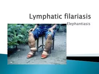
Chronic Manifestation Chronic (Obstructive) lesions takes 10-15 years. This is due to the permanent damage to the lymph vessels caused by the adult worms, the pathological changes causing dilation of the lymph vessels due to recurrent inflammatory episodes leading to endothelial proliferation and inflammatory granulomnatous reaction around the parasite. Initially, it starts with pitting oedema which gives rise to browny oedema leading to hardening he tissues. Still late, hyper pigmentation, caratosis, wart like lesions are developed. Eg. Hydrocele (40-60%), Elephantiasis of Scrotum, Penis, Leg, Arm, Vulva, Breast, Chyluria.
2. Occult Filariasis (TPE) • Occult or Cryptic filariasis, in classical clinical manifestation mf will be absent. Occult filariasis is believed to be the result of hyper responsiveness to filarial antigens derived from mf. Seen more in males. Patients present with paroxysmal cough and wheezing, low grade fever, scandy sputum with occasional haemoptysis, adenopathy and increased eosinophilia. X-ray shows diffused nodular mottling and interstial thickening.
Classification of Lymphoedema • Lymphoedema is classified into 7 stages on the basis of the presence & absence of the following: • Oedema • Folds • Knobs • Mossy foot • Disability
Stages of Lymphoedema of the Leg (Stage I) • Swelling reverses at night • Skin folds-Absent • Appearance of Skin-Smooth, Normal
Stages of Lymphoedema of the Leg (Stage II) • Swelling not reversible at night • Skin folds-Absent • Appearance of skin-Smooth, Normal
Stages of Lymphoedema of the Leg (Stage III) • Swelling not reversible at night • Skin folds-Shallow • Appearance of skin-Smooth, Normal
Stages of Lymphoedema of the Leg (Stage IV) • Swelling not reversible at night • Skin folds-Shallow • Appearance of skin - Irregular, • * Knobs, Nodules
Stages of Lymphoedema of the Leg (Stage V) • Swelling not reversible at night • Skin folds-Deep • Appearance of skin – Smooth or Irregular
Stages of Lymphoedema of the Leg (Stage VI) • Swelling not reversible at night • Skin folds-Absent, Shallow, Deep • Appearance of skin *Wart-like lesions on foot or top of the toes
Stages of Lymphoedema of the Leg (Stage VII) • Swelling not reversible at night • Skin folds-Deep • Appearance of skin-Irregular • Needs help for daily activities - Walking, bathing, using bathrooms, dependent on family or health care systems
Pathology of Lymphatic Filariasis • The pathology associated with lymphatic filariasis results from a complex interplay of the pathogenic potential of the parasite, the tissue response of the host and external bacterial and fungal infections. Most of the pathology associated with LF is limited to the lymphatics.
The damage to the lymphatic vessels is mediated both by an immune response to the adult worms as well as by a direct action of the parasite or the product released by them. In the absence of inflammation, marked lymphatic dilation with lymphoedema is seen in experimental animals with immune deficiency and when immuno competent cells are induced, it results inflammatory granuloma reactions around the parasite and subsequent obstructions of the lymphatic vessel occurs leading to lymphoedema.
Twin Pillars of Lymphatic Filariasis Elimination • Interrupt transmission • Control Morbidity (relief of suffering) # Community-level care of those with disease • Lymphoedema • Acute inflammatory attacks • Hydrocele repair
Management of Lymphatic Filariasis • Treating the infection • Treatment and prevention of Acute ADL attacks • Treatment and prevention of Lymphoedema
Treating the infection Remarkable advances in the treatment of LF have recently been achieved focusing not on individual but on community with infection, with the goal of reducing mf in the community, to levels below which successful transmission will not occur.
Chemotherapy of Filariasis Drugs effective against filarial parasites • Diethyl Carbomazine citrate (DEC) • Ivermectin • Albendazole • Couramin compound Treatment of microfilaraemic patients may prevent chronic obstructive disease and may be repeated every 6 months till mf and/or symptoms disappears.
Diethyl Carbomazine Citrate (Hetrazan, Banocide, Notezine) • Mode of action: DEC do not have direct action of parasite but mediate through host immune system. • Very effective against mf (Microfilariacidal) • Lowers mf level even in single dose • Effective against adult worms in 50% of patients in sensitive cases. • Dose: 6mg/Kg/12 days • Recent dosage: 6mg/Kg single dose • Adverse reactions are mostly due to the rapid destruction of mf which is characterised by fever, nausea, myalgia, sore throat, cough, headache. • No effect on the treatment of ADL • Drug of choice in the treatment of TPE.
Ivermectin • Mode of action: Directly acts on mf and no action on adults. • Very effective against mf (Microfilariacidal) • Lowers mf level even in single dose of 200µg – 400µg/Kg body weight • No action on TPE • Drug of choice in Co-endemic areas of Onchocerciasis with LF. • Adverse reactions are lesser but similar to that of DEC • Microfilariae reappears faster than DEC
Albendazole • This antihelmenthic kills adult worms • No action on microfilariae • Dose: 400mg/twice day /2 weeks • With combination of DEC & Ivermectin, it enhances the action of the drugs. • It induces severe adverse reactions in hydrocele cases due to the death of adult worms.