Download
1 / 45
450 likes | 628 Views
Respiratory disease in the newborn. Dr. Rozin Ilya. הצגת מיקרה. תינוק שנולד בשבוע 40 בניתוח קיסרי דחוף עקב חוסר התקדמות וניטור עם סימני סבל עוברי. אם עם חום סביב לידה עד 38.0. בלידה מים מקוניאליים, תינוק ללא נשימה ספונטאנית,רפוי,כחול.

E N D
Respiratory disease in the newborn Dr. Rozin Ilya
הצגת מיקרה • תינוק שנולד בשבוע 40 בניתוח קיסרי דחוף עקב חוסר התקדמות וניטור עם סימני סבל עוברי. • אם עם חום סביב לידה עד 38.0. • בלידה מים מקוניאליים, תינוק ללא נשימה ספונטאנית,רפוי,כחול. • בוצע אינטובציה וסקשיון מהקנה עם תוכן נוזל מקוניאלי. הנשמה למשך דקה עם הופעת נשימות ספונטאניות,עליה בטונוס.דופק טוב. • בוצע אקסטובציה עם נשימות עצמוניות וסימני מצוקה נשימתית קלה.קיבל חמצן סביבתי. • אפגר 8\9 (עקב צבע וטונוס) • תינוק הועבר למעקב במחלקת תינוקות.
הצגת מיקרה • במחלקה נזקק לחמצן בכיפה עד 35%.סימני מצוקה נשימתית שהלכה וגברה יחד עם עליה בתצרוכת חמצן ל 60% ובהמשך ל 100% עם ריווי חמצן בדם 90%-88%. • בוצעו בדיקות דם – בעבודה. גזים בדם עם pH7.19,pCO2 75,pO2 35, Bic19,BE-6. • צילום חזה עם Patch בעיקר בבסיסי הריאות יותר מימין עם ציור ריאתי מעוט וצל הלב תקין. • הוחל טיפול אנטיביוטי ומתן נוזלים I.V. • להמשך טיפול ובירור עובר למחלקת לטיפול נמרץ פגים.
הצגת מיקרה • הונשם מייד אחרי קבלתו עם שיפור ב CO2,אך O2 נשאר נמוך,למרות עליה בלחצי הנשמה ו 100% חמצן. SatO2 הייתה סביב 85%. • לצורך הערכה קרדיאלית בוצע ECHO לב עם מבנה תקין,נצפה PDA רחב עם דלף מימין לשמאל ו PFO. • לאור הממצאים הוחל מתן Nitric Oxide עד 20 ppm עם עליה ב SatO2 ל 95% וירידה בתצרוכת חמצן ל 45%. • בשל לחץ דם גבולי לגילו הוחל מתן Dopamine. • בהמשך חל שיפור הדרגתי,כעבור מספר ימים עבר אקסטובציה ונשאר עם חמצן סביבתי למשך 7 ימים.שחרור לבית במצב טוב.
Signs and symptoms • Cyanosis • Grunting • Nasal flaring • Retraction • Tachypnea • Decreased breath sounds with rales and / or rhonchi • Pallor • Apnea
Causes Central or peripheral nervous system hypoventilation: • Birth asphyxia • Intracranial hypertension, hemorrhage • Over sedation ( direct or through maternal rout ) • Diaphragm palsy • Neuromuscular disease • Seizure
Causes Respiratory disease: Upper airway: • Choanal atresia / stenosis • Pier Robin syndrome • Intrinsic airway obstruction ( laryngeal / bronchial / tracheal / stenosis ) • Extrinsic airway obstruction ( bronchogenic cyst, duplication cyst, vascular compression )
Causes Respiratory disease: Lower airway: - Respiratory distress syndrome - Transient tachypnea - Meconium aspiration - Pneumonia ( sepsis ) - Pneumothorax - Congenital diaphragmatic hernia - Pulmonary hypoplasia
Causes Cardiac right to left shunt: Abnormal connection ( pulmonary blood flow normal or increased ): - Transposition of great artery - Total anomalous pulmonary venous return - Truncus arterious - Hypoplastic left heart syndrome - Single ventricle or tricuspid atresia with VSD & without PS
Causes Cardiac right to left shunt: Obstructed pulmonary blood flow ( pulmonary blood flow decreased ): • Pulmonic atresia with intact ventricular septum • Tetralogy of Fallot • Tricuspid atresia • Single ventricle with Pulmonic stenosis • Ebstein malformation of the tricuspid valve • Persistent fetal circulation ( PPHN ) • Critical Pulmonic Stenosis with PFO or ASD
Causes Methemoglobinemia: - congenital ( hemoglobin M, methemoglobin reductase deficiency ) - Acquired ( nitrates, nitrites ) Other: - Hypoglycemia - Adrenogenital syndrome - Polycythemia - Blood loss
Transient tachypnea of newborn • Usually in normal preterm or term vaginal delivery or C/S • Early onset of tachypnea, retraction, cyanosis ( O2 < 40%) • Usually recover rapidly within 3 day • In auscultation – clear sound • Chest x- ray : prominent pulmonary vascular marking, fluid in the intralobar fissures, overaeration, flat diaphragms, rarely pleural effusion. • Secondary to slow absorption of fetal lung fluid resulting in decreased pulmonary compliance and tidal volume and increased dead space • Treatment is supportive
Meconium aspiration • Found in 10-15% of births • Usually occurs in term or post-term infants • Meconium aspiration pneumonia – in 5% • Require mechanical ventilation – 30% • Death 3-5% • Pathogenesis: - peripheral and proximal airway obstruction - inflammatory and chemical pneumonitis - remodeling of pulmonary vasculature - atelectasis > V / Q mismatch - air trapping > air leaks - persistent pulmonary hypertension - acidosis, hypoxemia, hypercapnea
Meconium aspiration • In clinical signs – respiratory distress, - tachypnea persistent from few days to several weeks, - hypoxia and metabolic acidosis. • In chest x-ray – overdistention, typical – patchy infiltrates, coarse streaking of both lung, signs of PPH • Therapy – supportive care ( mechanical ventilation, used of exogenous surfactant, ECMO ) • Prevention – for depressed infant – intubations with suction.
Persistent pulmonary hypertension of newborn • Occurs in term and post-term infants • Predisposition factors: - birth asphyxia, - meconium aspiration pneumonia, - early onset sepsis, - RDS, - hypoglycemia, polycythemia, - maternal use of NSAID (PDA closed) or SSRI, - pulmonary hypoplasia (result of diaphragmatic hernia), - oligohydramnios, - pleural effusion.
Persistent pulmonary hypertension of newborn • In pathophysiology – this is circulation with fetal pattern of right to left shunting through the PDA and Foramen Ovale after birth. • PPHN is often idiopathic. • Some infants have low plasma arginine and nitric oxide metabolite concentration and polymorphisms of the carbamoyl phosphate synthase gene – defect NO production. • Incidence: 1/500 – 1/1500 live birth. • Survival varies with underline diagnosis.
Persistent pulmonary hypertension of newborn • In clinical picture: - infant become ill in the delivery room or within first 12 hr - initial signs may be minimal • Diagnosis: - hypoxia unresponsive to 100% of oxygen - gradient pO2 between preductal and postductal site of blood sampling > 20 mmHg or SatO2 > 5% by pulse oxymetry. - by ECHO – right to left shunt ,tricuspid regurgitation. - x-ray chest • D.D. – cyanotic heart disease.
Persistent pulmonary hypertension of newborn • Treatment : - Correcting predisposition disease - Oxygen administration - Talazoline – non selective alpha-adrenergic antagonist - Hyperventilation ( pCO2 =25 mmHg with pH 7.50-7.55) - Sedation ( Fentanyl ) - paralytic drugs – controversial - Inotropic therapy - Nitric Oxide ET inhalation ( reduce ECMO by 40% ) - Prostacyclin (PGI 2) I.V.
Persistent pulmonary hypertension of newborn - Extracorporeal Membrane Oxygenation ( ECMO ) – is form of cardiopulmonary bypass that augments systemic perfusion and provides gas exchange. Criteria: - Oxygenation Index: (MAP * FiO2 * 100) / PaO2 (35-60) - Alveolar Arterial Oxygen Gradient: FiO2 (P-47) – PaO2 – PaCO2 [FiO2 + (1-FiO2) / R] P – barometric pressure(760), R – respiratory quotient(0.8) (> 605-620) - PaO2: < 40 mmHg - Acidosis and Shock: pH<7.25 or + hypotension
Congenital diaphragmatic hernia • May be due to defective formation of the pleuroperitoneal membrane. • Associated with pulmonary hypoplasia. • Incidence of CDH 1/2000 – 1/5000 live birth • Female : Male = 2 : 1 • Defect more common – left (85%) • Most common sporadic. • Associated anomalies in 30% (CNS lesion, Esophageal Artesia, omphalocele, CVS lesion) • Initial management – aggressive respiratory support with immediately intubation. Surfactant therapy commonly use, but no study for that is beneficial.
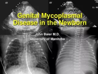
Pneumonia Bacterial infection is possible cause of neonatal respiratory distress. Common pathogens include: • group B streptococci (GBS), • Staphylococcus aureus, • Streptococcus pneumoniae, • gram-negative enteric rods. Pneumonia and sepsis have various manifestations, including the typical signs of distress as well as temperature instability.
Pneumonia Risk factors for pneumonia include: • prolonged rupture of membranes, - prematurity, • maternal fever. Prevention of GBS infection through screening and antepartum treatment reduces rates of early-onset disease including pneumonia and sepsis, by 80 percent. Intrapartum antibiotics at least four hours before delivery. Chest radiography helps in the diagnosis, with bilateral infiltrates suggesting in utero infection. Pleural effusions are present in 2/3 of cases. Serial blood cultures may be obtained to later identify an infecting organism.
Extrapulmonary air – leak syndrome Pneumothorax, defined as air in the pleural space, can be a cause of neonatal respiratory distress when pressure within the pulmonary space exceeds extrapleural pressure. It can occur spontaneously or as a result of infection, meconium aspiration, lung deformity, or ventilation barotrauma. The incidence of spontaneous pneumothorax is 1 to 2 percent in term births, but it increases to about 6 percent in premature births.
Extrapulmonary air – leak syndrome Pneumomediastinum occurs in at least 25% of patients with pneumothorax Usually asymptomatic Subcutaneous emphysema often asymptomatic and pathognomonic of pneumomediastinum If trapped air is great – neck veins are distended and - blood pressure is low it’s result of tamponade of the systemic and pulmonary vein.
Extrapulmonary air – leak syndrome Pulmonary interstitial emphysema (PIE) may: - precede the development of a pneumothorax - occur independently In pathogenesis: - increased alveolar-arterial oxygen gradient - increased intrapulmonary shunting - progressive enlargement of blebs of air may result in cystic dilatation. In therapy with oxygen and high frequency ventilation
Differential diagnosis with cyanotic CHD • Central cyanosis • Lack or minimal respiratory distress signs • Systolic murmur • Evaluation by ECHO • Chest x-ray • Hyperoxic test
Hyperoxic test • Placing in 100% oxygen concentration • During for 5 to 10 minutes • Sampling arterial gas or monitoring oxygenation non invasively • If PaO2 level higher than 100 mmHg - good • If PaO2 level above 40-50 mmHg – sign to right to left shunting
Evaluation and first line therapy a child with cyanosis • Anamnesis • Clinical signs and symptoms • Oxygen therapy • Blood gas measurement • CBC and blood culture • Chest x-ray • ECG if need • NPO • Fluid intravenously • Stomach decompression • Mechanical ventilation if need