Download
1 / 65
670 likes | 943 Views
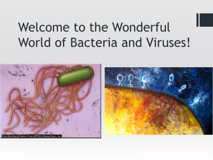
Welcome to the Wonderful World of Bacteria and Viruses!. What are Microbes?. Microbes are living things that are too small to be seen with the unaided eye They include bacteria, fungi, protozoa and microscopic algae.

E N D
What are Microbes? • Microbes are living things that are too small to be seen with the unaided eye • They include bacteria, fungi, protozoa and microscopic algae. • Typically we view microbes as harmful, causing diseases, but only a minority of all microbes is pathogenic (disease-producing).
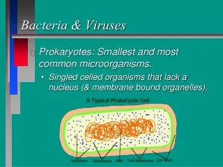
Bacteria • Bacteria are microbes, which are single-celled organisms that are too small to be seen without a microscope. • They are considered to be prokaryotes, which means that their DNA is not enclosed by a membrane. • Eukaryotic cells have a nucleus with a membrane. • Where can bacteria be found?
Bacterial Cell Parts • Capsule – outer covering on some bacteria; protects the cell and makes it more pathogenic; considered a glycocalyx or sugar coat • Cell wall – semi-rigid structure that maintains the shape of the cell and protects the interior of the cell from changes in the outside environment • The bacterial cell wall contains peptidoglycan. Some bacteria have just a few layers of peptidoglycan while others have many. • Peptidoglycan is not found anywhere else in the biological world.
Bacterial Cell Parts • Plasma membrane - thin structure inside the cell wall that surrounds the cytoplasm, serves as a selective barrier through which materials can enter and exit • Ribosomes - appear as small dots in the cytoplasm; they make proteins • Nucleoid – the area where the DNA is found in the cell • Bacterial chromosome- the long, continuous and usually circular double strand of DNA
Bacterial Cell Parts • Plasmids - small circular strands of DNA not connected to the main bacterial chromosome • Cytoplasm - everything inside the plasma membrane; mostly made up of water • Pili – hair-like structures that allow bacteria to attach to other cells and transfer DNA (conjugation) • Flagella - long tails that help bacteria move
Shapes of Bacteria • Come in three main shapes • Bacillus (rod) • Coccus (sphere) • Spirillum (spiral)
Arrangements of Bacteria • Straight chains of bacteria are referred to as strepto- • Two cells of bacteria are called diplo- • A cluster of bacterial cells is known as staphylo-
Gram Stain • Developed by the Danish bacteriologist Hans Christian Gram in 1884 • Classifies bacteria into two major groups: • Gram-positive • Gram-negative • Procedure • 1. Primary stain of crystal violet • 2. Mordant of Gram’s iodine • 3. Decolorizing agent of alcohol • 4. Counterstain of safranin
Gram Stain • The crystal violet and iodine combine within the cytoplasm of the bacterial cells. • Gram-positive - The color does not get washed out from the alcohol wash and the cells remain purple. • Have thicker layer of peptidoglycan in cell walls that enables the cell to retain the crystal violet • Gram-negative - The color is washed out by the alcohol and is no longer visible. When the counterstain safraninis applied, the cells appear pink. • The crystal violet can be washed out because they have a thinner layer of peptidoglycan
Gram Stain Gram-positive Gram-negative
Normal Flora/Microbiota • We all live with a variety of microbes on and in us. • These microbes make up our normal flora and include fungi, protists, but mostly bacteria. • Our body is made up of 1013 cells and we harbor about 1014 bacteria. • 10 times more bacterial cells than human cells! • They are in most cases beneficial to us because they protect our bodies from diseases by preventing the overgrowth of harmful microbes.
Normal Flora/Microbiota • Normal flora can be found throughout our body localized in certain regions. • Skin • Eyes (conjunctiva) • Nose and throat (upper respiratory system) • Mouth • Large intestine • Urinary system • Reproductive system
Factors that influence where normal flora/microbiotaare found • Availability of nutrients • Chemical and physical factors • Defenses of the host • Age • Gender
Normal Flora/Microbiota of Skin • Microbes on our skin must be able to withstand secretions from the sweat and sebaceous glands that have antimicrobial properties. • Keratin, a protein found on the skin, acts as a resistant barrier and the low pH of the skin can also inhibit many microbes. • These factors prevent the microbes that are in direct contact with the skin from becoming permanent residents.
Staphylococcus epidermidis • S. epidermidisis the most abundant inhabitant of the skin, especially the upper body.
Staphylococcus aureus • The nose is one of the most common sites for S. aureus. • It is a leading cause of bacterial disease in humans. It can be transmitted from the nasal membranes of a carrier to a susceptible host (immunocompromised).
Propionibacterium acnes • Located on greasy areas of the skin, such as the forehead • Can become trapped in hair follicles and cause inflammation and acne • Different species of Propionibacterium can live on the sides of our nose and on our armpits.
Symbiosis • The relationship between the normal flora and the host is called symbiosis. • Symbiosis - living together • Three types of symbiotic relationships • 1. mutualism • 2. commensalism • 3. parasitism • Animal symbiosis (4:20)
Mutualism • Where both the host and bacteria are thought to derive benefits from each other, it is referred to as being mutualistic. • Example: E. coli synthesizes vitamin K and some B vitamins that are absorbed into our blood stream. The large intestine, in return, provides nutrients needed by the bacteria.
Commensalism • Commensalism - where one organism benefits and the other is unaffected • Many of the bacteria that make up our normal flora are commensals. • These include the bacteria that are on the surface of our eyes and skin and some bacteria in the ear that live on secretions and sloughed-off cells.
Parasitism • Parasitism -when one organism benefits at the expense of the other organism • In some situations normal flora can make us sick. This can occur when the normal flora leave their habitat. • Example: When E. coli leave their habitat of the large intestine and gain access to other body parts, such as the urinary bladder, lungs or spinal cord, they can cause infections such as urinary tract infections, pulmonary infections, meningitis and abscesses.
Nutrients • Bacteria can colonize only where they can be supplied the right nutrients. • These nutrients can come from excretory products of the cells, substances in body fluids and dead cells, and food in the gastrointestinal tract.
Chemical and Physical Factors that Affect Bacterial Growth • Temperature • pH • Available oxygen and carbon dioxide • Salinity • Sunlight (UV rays)
Mechanical Factors that Affect Bacterial Growth • Certain areas of the body have different mechanical factors that can affect normal flora from colonizing. • These include: • Chewing action of teeth • Tongue movements • Flow of saliva and digestive secretions in gastrointestinal tract • Muscular movements of throat, stomach, intestines • These mechanical factors can trap or dislodge microbes.
How do Bacteria Make us Sick? • It’s not the bacteria themselves that make us sick; it’s what they produce that makes us sick. • Bacteria produce substances called toxins that cause illness and disease. • There are two types of toxins: exotoxins and endotoxins • Exotoxins are proteins that are secreted by bacteria and are products of their metabolism. • Generally produced by Gram-positive bacteria • Some of the deadliest substances known (C. botulinumtoxin) • Disease examples: food “poisoning”, TSS, tetanus, botulism, diphtheria, gas gangrene • Endotoxins are components of the bacterial cell wall (lipopolysaccharides). • Generally found in Gram-negative bacteria • Disease examples: no specific diseases; cause fever, hemorrhaging, shock, miscarriage, coma, death
Defenses of Host • The body has many defenses against microbes that include: • Activated cells that kill microbes • Cells that inhibit growth • Prevent adhesion to cell surfaces • Neutralize toxins that microbes produce • These defense mechanisms are helpful in fighting pathogens. • However, their role in regulating normal flora is unclear.
Biofilms • Masses of microbes attached to a surface; increases pathogenicity (involved in 70% of infections) • The “slime” is made primarily of polysaccharides with some DNA and proteins. • The microbes are connected to each other, enabling them to chemically communicate with each other, transfer DNA, and share nutrients. Plaque
Biofilms • Because of the complex chemical composition of the slime, biofilms are resistant to immune systems, disinfectants, and antibiotics (about 1000 times more resistant). • Significant problem on internal medical devices such as catheters, stents, and mechanical heart valves; also problematic on contact lenses • When microbes grow in biofilms, they communicate with each other. • Examples: plaque, pool algae, shower scum • Bacterial communication (18:00)
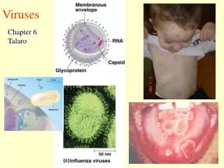
Virus Characteristics • Viruses contain DNA or RNA, but not both • Possess a protein coat • Some viruses have surface spikes • Most viruses infect only specific types of cells in one host • Host range is determined by specific host attachment sites and cellular factors • First isolated and studied in 1935 (tobacco mosaic virus) • Phage therapy and oncolytic viruses
Are Viruses Alive?? • Viruses are inert outside a host cell = nonliving • They have few or no enzymes of their own, so they need their host cells’ enzymes. • However, once they enter a cell, their nucleic acids become active and they start multiplying. • They cause infection and disease just like living pathogens. • They could be considered: • a very complex mixture of chemicals, or • a very simple living organism
Viral Structure • Virion - a complete, fully-developed, infectious viral particle composed of nucleic acid and surrounded by a protective protein coat (capsid) • Capsomere – protein subunit of a capsid; can be used to identify a particular virus • Envelope – lipid, protein, and carb complex covering capsule; many times contains parts of host membrane • Used to help virus enter host cell • Spike – carb-protein complex projecting from envelope; can be used to ID virus • Used to attach virus to host cell and clump cells together
How do Viruses Reinfect? • Why do we get influenza more than once? • The surface proteins (spikes) of many viruses, like influenza, can easily mutate. • Although you may have produced antibodies to one influenza virus, the virus can mutate and infect you again. Influenza viruses – (notice the spikes) Virus video (3:38)
Multiplication of Bacteriophages (Lytic Cycle) • A virus must invade and take over a host cell in order to multiply; it needs the host cell’s enzymes and nucleic acids. • Attachment Phage attaches by tail fibers to host cell receptors • Penetration Phage lysozyme (enzyme) opens cell wall; DNA is injected into cell • Biosynthesis Production of phage DNA and proteins; no complete phages yet; step when host cell is taken over • Maturation Assembly of phage • Release Phage lysozyme breaks cell wall and “baby” phages are released to wreak more havoc
Attachment Lytic Cycle of a Bacteriophage (a) Penetration Biosynthesis
Lytic Cycle of a Bacteriophage (b) Maturation Release
Lytic/Lysogenic Cycles • Lytic cycle: Phage causes lysis and death of host cell • Lysogenic cycle: Prophage DNA (inserted phage DNA) incorporated in host DNA • Host cell does not die • Every time host divides, so does the prophage DNA. Lysis of E. coli
Viruses and Cancer • Almost anything that alters cellular DNA (oncogenes) has the potential to make normal cells cancerous. • Several types of cancer are known to be caused by viruses (~10% of all cancers). • Relationship between viruses and cancer first demonstrated in 1908 (chicken leukemia) • Oncoviruses – viruses that transform normal cells into cancerous cells. • Transformed cells have increased growth, loss of contact inhibition, and viral-specific T antigens (antigens that disrupt the normal cell cycle). • The genetic material of oncogenic viruses becomes integrated into the host cell's DNA (similar to lysogeny).
Latent/Persistent Viral Infections • Latent Viral Infections • Virus remains in asymptomatic host cell for long periods • Cold sores, shingles, oncogenic viruses • Many remain latent in nervous tissue • Can be reactivated by immunosuppression • Persistent Viral Infections • Disease progresses over a long period; generally fatal; disease presents years later • Often caused by another virus • Measles and fatal encephalitis; HBV and liver cancer; human papillomavirus and cervical cancer
Plant Viruses and Viroids • Plants are generally protected by cell wall • Plant viruses enter through wounds or via insects that suck plant’s sap • Viruses can multiply inside insect cells, then get passed to plant • Viroids • Viroids are infectious pieces of RNA; potato spindle tuber disease • Responsible for millions of $ in crop damage tobacco mosaic virus
Prions • Infectious proteins first explained in 1982 • Inherited and transmissible by ingestion, transplant, and surgical instruments; can be spontaneous as well • Treated with proteases or NaOH and autoclave • Most resistant to chemical biocides because when altered, they’re extremely stable and resistant to denaturation • Many are believed to be inherited (the case with the five human prion diseases) • PrPC: normal cellular prion protein; on cell surface • PrPSc: scrapie protein; accumulates in brain cells forming plaques (plaques used for postmortem diagnosis; don’t appear to be cause of cell death)
Prions Microscopic "holes" are characteristic in prion- affected brain tissue sections, causing the tissue to develop a "spongy" appearance.