Download
1 / 30
300 likes | 407 Views
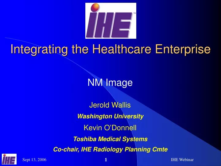
Integrating the Healthcare Enterprise. NM Image. Jerold Wallis Washington University Kevin O’Donnell Toshiba Medical Systems Co-chair, IHE Radiology Planning Cmte. NM Image Transaction Diagram. Evidence Creator. Image Display. Image Archive. Image Manager. Acquisition Modality.

E N D
Integrating the Healthcare Enterprise NM Image Jerold Wallis Washington University Kevin O’Donnell Toshiba Medical Systems Co-chair, IHE Radiology Planning Cmte IHE Webinar
NM ImageTransaction Diagram EvidenceCreator ImageDisplay ImageArchive ImageManager AcquisitionModality Vol 1- 16.1 Actors and Transactions in NM Profile IHE Webinar
NM ImageTransaction Diagram EvidenceCreator ImageDisplay ImageArchive ImageManager AcquisitionModality IHE Webinar
Acquisition Modalities • Create NM objects as specified (Series, attribute & vector handling) • Image orientation • Patient orientation • Include specific codes in Cardiac studies • Patient state codes • Reorientated cardiac image codes • Slice progression direction • 4.8.4.1.2.2 Storage of NM Images (NM) • Also see revisions of table 4.8-2 in CP IHE Webinar
Table 4.8-2 (Revised) IHE Webinar
NM ImageTransaction Diagram EvidenceCreator ImageDisplay ImageArchive ImageManager AcquisitionModality IHE Webinar
Evidence Creators • Create NM objects as specified (Series, attribute & vector handling) • Include specific codes in Cardiac studies • 4.8.4.1.2.2 Storage of NM Images (NM) • Also see revisions of table 4.8-2 in CP IHE Webinar
Result Screen Export Option IHE Webinar
Result Screen Export Option:Evidence Creators • Render Dynamic Result Screens • Multi-Frame Grayscale Byte Secondary Capture • Multi-Frame True Color Secondary Capture • Creating multiple single frame objects is NOT sufficient. • Render Static Result Screens • Using single frame DICOM Secondary Captures is sufficient • Using Multi-frame to render single or several related static result screens is recommended • Must generate appropriate Secondary Capture objects from any Result Screens created. • 4.18.4.1.2.4 Result Screen Export Option IHE Webinar
NM ImageTransaction Diagram EvidenceCreator ImageDisplay ImageArchive ImageManager AcquisitionModality IHE Webinar
Image Manager/Archives • Store/Retrieve NM Objects • Store/Retrieve Secondary Capture (SC & MFSC) Objects • Must not strip private elements • Support Query on Study Description • Must return Series Description when requested • 4.8.4.1.3 Expected Actions • 4.18.4.1.3.1 DICOM Image Storage SOP Classes • Table 4.14-1. Images Query Matching and Return Keys IHE Webinar
NM ImageTransaction Diagram EvidenceCreator ImageDisplay ImageArchive ImageManager AcquisitionModality IHE Webinar
Image Display • Display NM and Result-Screen Images • Support specified NM essential display capabilities • Additional attribute handling, with display of values or use as indicated • e.g. Patient State, Image Orientation, Detector Sequence, View Code Sequence • 4.16.4.2.2.3 Display of NM Images • Revision in CP IHE Webinar
Understood by dedicated NM systems Often Misunderstood by general storage and display systems Multiframe Dynamic Anterior Posterior Multi-energy NM Image Format NM Image • Image vectors Volume 1 – E.4 IHE Webinar
General NM Option Whole body display Cardiac NM Option ACC NM Cardiac display Options: Image Displays Displays must support at least one of these options! In CP 4.16.4.2.2.3.6 General NM Option 4.16.4.2.2.3.7 Cardiac NM Option IHE Webinar
Image Display • Handle multi-frame / multi-vector images • Multiple simultaneous cines • Grayscale and color result screens • Dynamic and static result screens • Original resolution and scaled • Separate adjustment of upper and lower window levels • Apply local color tables to grayscale screens • Add new local color tables to system • Change intensity of grayscale result screens • Display Series Description tag • Display orientation labels IHE Webinar
Upper and Lower Levels • Upper and lower window levels • (Not just center/width) • Separate • Design is up to you IHE Webinar
Image Display • Allow user to select different vectors/framesets of complex multi-frame images • Allow user to separately adjust intensity for different framesets (eg, phases) in the same “image” IHE Webinar
Multiple Cines IHE Webinar
Display Formats A1 A2 A3 A4 A5 A6 A7 A8 A9 … … … … … … … … … … … … … … … … … … … … … … A32 … A1 A2 B1 B2 C1 C1 • Grid display • Frames from one image set in a grid • Fit display • Frames from multiple image sets in a grid • Comparison Display removed in CP IHE Webinar
Table 4.16-1 (Revised) IHE Webinar
Table 4.16-1 (Revised) • Revised table in CP IHE Webinar
ACC NM Cardiac Display • Generate long axis from short axis images • Display of 2 image sets (Stress and Rest) • Use patient state tags to set display order • Follow ACC guidelines for display • Controls for adjusting stress and rest • Image alignment • Window upper and lower levels. • Description in CP IHE Webinar
Short axis ACC NM Display Example Only Short axis data supplied • Vertical Long • Horizontal Long IHE Webinar
Short ACC NM Display Example Stress Rest Upper/lowerAdjustments Stress Rest Stress Rest • Vertical Stress Rest • Horizontal Stress Rest IHE Webinar
Another ACC Display Example • Different Layout • Same stress and rest views IHE Webinar
MPR Option: Image Displays • Adds Image Display requirements to support Review of SPECT NM Images with Multi-planar reconstruction (MPR) • Transaxial • Sagittal • Coronal • Described in CP IHE Webinar
Points of Interest - NM • Appendix E, Volume 1 (Informative) • New to Nuclear Medicine? • Trouble matching NM to Scheduled Workflow? • Start Here. • E.2 NM Workflow – What happens in NM procedures • E.3 NM Worklists – How to schedule/model it • E.4 NM Data – What to expect in the DICOM • E.5 NM Display – How it should be presented IHE Webinar
Questions ? • This document is at • http://gamma.wustl.edu/ihe • (Also is on the IHE FTP server) • Links to the Framework and CP at • www.snm.org/ihe IHE Webinar
NM ImageStandards Used • DICOM NM Image Object • DICOM Multiframe Secondary Capture • Dynamic Results • Collection of Static Results • DICOM Secondary Capture • Static Results • ACC NM Cardiac Display format IHE Webinar