Download
1 / 60
600 likes | 744 Views
Circulatory System. 10.2. is located in the thoracic cavity behind the sternum it is about the size of your fist It uses up 15% of the bodies O2 and food supply it is surrounded by a fluid-filled membrane called the pericardium which prevents friction. 10.2 The Heart.

E N D
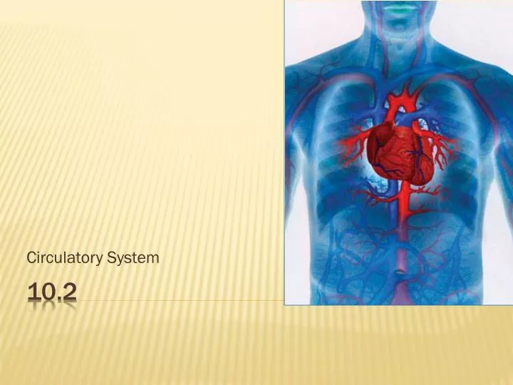
Circulatory System 10.2
is located in the thoracic cavity behind the sternum it is about the size of your fist It uses up 15% of the bodies O2 and food supply it is surrounded by a fluid-filled membrane called the pericardium which prevents friction 10.2 The Heart
cardiac muscle is very strong a layer of fat protects and cushions the heart the coronary artery supplies the heart with blood
The heart has 4 chambers • 2 atria (L&R) • 2 ventricles (L&R) • The Septum is a wall of muscle separates the right and left sides of the heart
atria act like holding chambers for blood entering the heart • blood from the pulmonary system enters the left atrium • blood from the systemic system enters the right atrium Atria
ventricles are strong muscular chambers that pump blood away from the heart Ventricles
Your Assignment: Heart Diagram – label the structures (see page 320) aorta left pulmonary artery septum left pulmonary vein right atrium right pulmonary artery left atrium right pulmonary vein right ventricle 2 semilunar valves left ventricle 2 atrioventricular (AV) valves superior vena cava inferior vena cava chordae tendonae (draw this in)
pulmonary circulation = blood vessels that carry blood to and from lungs systemic circulation = blood vessels that carry blood to and from body A. One Way Blood Flow
Superior vena cava= carries deoxygenated blood from your head to your heart Inferior vena cava= carries deoxygenated blood from your body to your heart Two Largest Veins:
Veins carry blood to the heart • deoxygenated blood reaches the heart through inferior and superior vena cava and empties into the right atrium • blood moves through atrioventricular or (AV) valve into right ventricle (***note: these are supported by bands of tendons called chordae tendinae) Blood Flow
Blood is pumped through semilunar valve into the left and right pulmonary arteries (***note: these are the only arteries that carry deoxygenated blood)
In lungs, oxygen diffuses into the blood Oxygenated blood enters pulmonary veins which take it back to the heart (***note: these are the only veins that carry oxygenated blood)
Blood enters left atrium then moves through atrioventricular or (AV) valveinto the left ventricle oxygenated blood is pumped through semilunar valve into the aorta (largest artery) where it travels to body tissues (*** note: these valves prevent the backflow of blood from the artery into a ventricle)
tissues use oxygen/nutrients/fluids that is in blood • then deoxygenated blood moves through vein system into inferior and superior vena cava
coronary arteries supply the heart muscle cells with O2 and nutrients Is chest pain (angina) that occurs when too little O2 reaches the heart Angina
it could be but not always caused by a blockage if it is a blockage, then the blockage can be bypassed by using veins from other parts of the body …these are grafted into the heart
Your Assignment: Draw red (oxygenated) and blue (deoxygenated) arrows on heart diagram to show blood flow into and out of heart
It is a technique used to detect coronary artery blockage B .Cardiac Catheterization
A catheter (small thin hollow tube) is passed into an artery in the groin The catheter is then pushed up through the aorta and into the heat
A dye is then injected into the catheter The dye travels through the blood vessels while its image is traced by a fluoroscope
An area of restricted blood flow pinpoints the region of blockage Figure 5 Page 323 Blood samples can also be taken to determine how much O2 is in the blood in different chambers of the heart
Cardiac muscle tissue is the only muscle that can contract without external nerve stimulation Muscle with this ability is called myogenic muscle C. Setting the Heart's Tempo
http://www.nhlbi.nih.gov/health/dci/Diseases/hhw/hhw_electrical.htmlhttp://www.nhlbi.nih.gov/health/dci/Diseases/hhw/hhw_electrical.html
The heart's tempo is set by the sinoatrial or (SA) node This is a bundle of specialized nerves and muscle located in the upper right atrium The SA nodes acts as a pacemaker, and sets a rhythm of about 70 beats/min
The contraction is generated in the SA node electrical impulses pass on to both atria, causing them to simultaneously contract Theimpulses then move to the atrioventricular node (AV node) (which acts like a conductor passing the nerve impulses) Nervous Control of the heart
The message is then relayed quickly down to special nerves in the septum called the Bundle of His It is then sent to the Perkinje Fibres The result is both ventricles are now stimulated to contract at the same time
Monitor the concentration of chemicals in the blood and blood pressure: • baroreceptors detects blood pressure in the aorta and carotid artery • chemoreceptors detects amount of CO2 or O2 in blood Receptors
these receptors signal the medulla oblongata the medulla oblongata responds by stimulating the autonomic nervous system
Consists of the: • parasympathetic and sympathetic nervous system • regulates equilibrium • its nerve impulses can affect heart rate Autonomic nervous system
parasympathetic nervous system: tells heart to beat at a normal rate sympathetic nervous system: tells heart to increase heart rate Parasympathetic Vs. Sympathetic
sympathetic nerves send impulses to the pacemaker to increase heart rate this increases blood flow to the tissues Heart rate exceeding 100 beats/minute = tachychardia E.g. exercise, drugs (cocaine, caffeine) Stress
parasympathetic nerves are stimulated A nerve impulse is sent to the pacemaker to slow the heart down Low heart rate = bradycardia Relaxation
an electrocardiogram (ECG) measures the electrical activity of the heart changes in the electrical current reveal normal or abnormal events in the cardiac cycle Electrical Conductivity of the Heart
Pwave = atrial contraction QRS = ventricular contraction T = ventricles relaxed Electrical Current (mV) Time (s)
By comparing electrocardiograph tracings doctors can determine areas of the heart that are having problems
http://www.nhlbi.nih.gov/health/dci/Diseases/hhw/hhw_pumping.htmlhttp://www.nhlbi.nih.gov/health/dci/Diseases/hhw/hhw_pumping.html