Download
1 / 52
530 likes | 682 Views
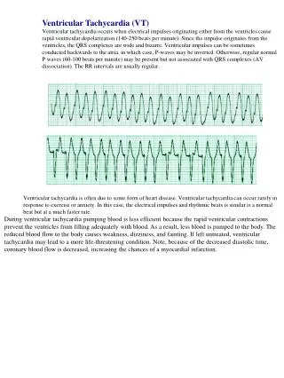
Ventricular tachycardia in abnormal heart. Dolly mathew. VT after MI . Sustained monomorphic VT- 3% extensive MI LV dysfunction LV aneurysm septal involvement Successful revascularization - <1%. pathophysiology.

E N D
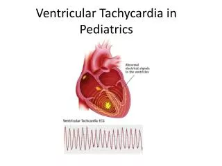
Ventricular tachycardia in abnormal heart Dolly mathew
VT after MI • Sustained monomorphic VT- 3% extensive MI LV dysfunction LV aneurysm septal involvement • Successful revascularization - <1%
pathophysiology • Anatomic substrate – extensive scar • Arises from surviving myocytes • Healthy & damaged myocardium interlaced with the fibrous scar at border zone of scar • Conduction is slow & discontinuous due to fibrosis & abnormalities in gap junctions • gradually develops in the first 2 weeks after myocardial infarction • remain indefinitely
Triggers – a/c ischemia - surges in the autonomic tone - heart failure • Once sustained monomorphic VT occurs, risk continues indefinitely, even if a/c ischemia & heart failure adequately controlled
Substrate modified by ischemic insults late ventricular remodelling worsening pump function Neurohormonal activation progressive LV dilatation increase in wall tension
Mechanism of ischemic VT • Reentry – macro/ micro reentry • Repolarization of individual myocardial cells not homogenous • Some cells excitable, some refractory
Sinus rhythm mapping in a patient with VT in the setting of extensive healed AWMI red ( dense scar) = 0.5 mV purple = 1.5 Mv intervening colors represent voltage values in between
In MI • Mostly from LV / septum • VT - LV apex – RBBB - rt superior axis • VT - upper half of septum - LBBB - rtinf axis • anterosupr LV – RBBB – Rtinf axis • post inf LV – RBBB – Lt supr axis
QRS morphology • The more rapid the initial forces, the more likely VT arising from normal myocardium (Josephson & Callans;heart rhythm2004) • Slurring of the initial forces – scarring • low amplitude VT – diseased myocardium • Notching of the QRS – scar • qRr, qr, QR complexes – s/o infarct • Septal VT less wide QRS
ECG features • 12 lead ECGs of 297 LBBB monomorphic VT recorded during catheter ablation ; 95 scar VT , 23 idiopathic • Diagnosis of scar based on SR ECG, cardiovascular imaging, & catheter mapping • Precordial transition beyond v4, notching of S downstroke in v1/v2 , onset of QRS- S nadir v1 >90 ms , were independent predictors of scar related VT • scar VT if any of the above criteria met • Idiopathic if none • In prospective validation,this algorithm was highly sensitive (96%) & specific (83%) for scar LBBB VTs (Adrianus P, Wijnmaalen et al,Circulation may 2011)
Sustained Ventricular Tachycardia:Role of the 12-lead Electrocardiogramin Localizing Site of OriginMARK E. JOSEPHSON, M.D., LEONARD N. HOROWITZ, CIRCULATION 1981 • QRS morphology of 41 morphologically distinct VT was correlated with their site of origin as determined by catheter and intraoperative mapping. • 12-lead ECG could not precisely identify the site of origin in patients with CAD • Could differentiate anterior from posterobasal regions, particularly in VT -LBBB. • ECG was less useful in localizing VT-RBBB because of overlapping patterns • General QRS patterns were useful in differentiating anterior from posterior regions of origin
ECG pattern less likely to predict site of origin in AWMI than with IWMI (37% vs 74% ; p< 0.001) • VT-LBBB on or adjacent to septum • VT-RBBB septal/ free wall location ( 73 vs 31% ; p< 0.001) • Relationship between the 12-lead ECG during VT and endocardial site of origin in patients with coronary artery disease;JM Miller ;Circulation 1988;,
The QRS morphology in post-MI VT, study of 100 tracings compared with 70 cases of idiopathic VT ( P. COUMEl, J. F. LECLERCQ, P. ATTUEL and P. MAISONBLANCHE) • The two groups of tracings differed in terms of QRS axis, most often normal in idiopathic VT (75%) and abnormal in MIVT (74%) • The sum of QRS amplitude in unipolar limb leads was greater in idiopathic VT (4.3±1.3 mv, mean±S.D.) than in MIVT (2.6±0.8 mv, P>0.001) • The QRS width was also different: 135±11 ms in idiopathic VT vs. 171±32 ms in MIVT (P>0.001) • The QRS morphology in MIVT- QR pattern in leads other than aVR, or a QS pattern in V5–V6 • These two aspects were constantly absent in idiopathic VT & present in 89%of MIVT • ECG signs of MI observed in the same leads during sinus rhythm and during VT, In only 38 MIVT tracings • In 51 MIVT tracings the location of the MI indicated by the VT tracing differed from that displayed in sinus rhythm
Clinical presentation & mgt • Determinants of hemodynamic stability- rate, LV fn, ischemia, MR Sedation, i/v medicines, DC cardioversion
Long term mgt • Goal of longterm therapy-a) pvt of SCD b) Rec of symp VT • Asymptomatic NSVT in pts with NLVEF- no treatment • Symptomatic NSVT in pts with NLVEF- betablockers • Cardiac arrest survivors / SUS VT in ↓LVEF- ICD • PRIMARY PVT - ICD > AMIOD- pvt of SCD • SECONDARY PVT - Class lll > l - ICD > amio in LVEF<35% • CAD-NL LVEF + SUST VT - amio, icd + amio, RFA
subendocardial resection of arrhythmogenic focus • Cryoablation • Laser vaporization • Photocoagulation
Ventricular arrhythmias in the setting of coronary artery disease all available antiarrhythmics except Amiodarone, l-Sotaloland Dofetilide increase mortality in the post MI population
Secondary Prevention of SCD survivors of card arrest or sustained VT- ICD provides the lowest mortality.
Primary Prevention of SCD in Ventricular Arrhythmias a prior MI, decEF and NSVT -ICD provides the lowest mortality.
Primary Prevention of SCD in absence of Ventricular Arrhythmias patients with significant LV dysfunction - best survival with ICD
VT in non ischemic cardiomyopathyDCM • Asymptomatic VT common • Incidence – 50-60% DCM, resp for 8-50% deaths • Factors contributing- -myocardial fibrosis, scar -increased circulating catecholamines -increased sympathetic tone -stretch induced afterdepolarizations -Sustained stress induced shortening of refractory period reentry
Pathophysiology - subendocardial scarring 30% (autopsy), 57% (histology) - Patchy fibrosis intermingled with viable myocardium – substrate for reentry - Basal & mid myocardial LV • mechanism - Macro reentry dominant mechanism - BBRVT- most characteristic - 6% vt in all patients, 41% in DCM
Severity of LV dysfunction most impt predictor of mortality • Association between QRS prolongation & mortality ( vesnarinone trial ) • ACEI – reduction in SCD due to VT, less frequent at 3 months (37% vs 46%); new VT less , at 1,2 yrs in enalapril group (VHeFT-II trial)
Beta blocker therapy relative risk reduction
Amiodarone - Used only on specific arrhythmic indications - Reduces ICD shock frequency , without worsening heart failure (SCDHeFT) • Biventricular pacing- severe drug refractory heart failure , in elderly • ICD- arrhythmic mortality reduction greater in classiii>ii ( DFINITE TRIAL) -No difference in mortality ( amiovs ICD) - Significant reduction in total mortality in icd group(SCDHeF) • LV assist devices – some pts tolerate ventricular arrhythmias well • Catheter ablation- failure due to mid myocardial source, critical isthmus, difficult epicardial access
VT in HOCM • SCD in adults with HCM- 1% NSVT – 8% • Amiodarone improve survival, young pts ( retrospective non randomized trials) • ICD implantation is reasonable for patients who have 1 or more major risk factor for SCD. (Level of Evidence: C)
No randomized trials regarding ICD therapy • Recom for life threatening VT/VF • Pts who have either one of the preceding life threatening arrhythmias or 1 or more other risk factors for SCD -NSVT,FH of premature SCD, unexplained syncope, LV thickness >30mm, abn exercise BP
Bundle Branch Re-Entry Ventricular Tachycardia • Macro re-entrant circuit employing • HPS • Both bundle branches • Ramifications of the left bundle • Transeptal myocardium • Hallmark: His-Purkinje system disease – functional or structural • very fast conduction velocity and a long refractory period
BBR -LBBB -antegrade direction -RB & reterograde LB BBR –RBBB- antegrade direction-LB & reterograde RB
His Catheter RB Catheter LB Catheter V Catheter
Surface ECG in sinus rhythm - non-specific or typical bundle branch block patterns with prolonged QRS duration • Total interruption of conduction in one of the BB would theoretically prevent occurrence of reentry • Can occur in patients with relatively narrow QRS complex -functional conduction delay
presyncope, syncope or sudden death - VT with fast rates > 200 bpm • LBBB pattern-mc VT morphology • VT of myocardial origin vs BBR-LB pattern – rapid intrinsicoid deflection initial ventricular activation through the HPS
BBRVT 1) Sinus rhythm – prolonged HV- prerequisite 2) Every ventricular depolarisationpreceeded by His deflections 3) HV interval during tachy ≥ HV interval of the spontaneous normally conducted QRS complexes 4) Documentation of H- RB – V – LB – VT LBBB H- LB –V – RB – VT RBBB
INTERFASCICULAR REENTRY TACHYCARDIA • usually has RBBB morphology • Antegrade - LAF & retro – LPF –RAD • Antegrade- LPF & retro – LAF- LAD
INTERFAS VT Vs RBBB RE ENTRY • HV interval shorter than sinus rhythm • LB potential before HIS deflections
High recurrence rate after drugs • RFA - first line therapy • choice is ablation of the RB
VT ARVD • Ventricular arrhythmias are usually exercise-related • sensitive to catecholamines. • right axis deviation, Supr axis ,LBBB morph in v1 • Multiple morphologies of ventricular tachycardia • multiple foci or pathways.
ARVC High Risk Features • Younger patients • Recurrent syncope • History of cardiac arrest or sustained VT • Clinical signs of RV failure or LV involvement • Patients with or having a family member with the high risk ARVD gene (ARVD2) • Increase in QRS dispersion ≥ 40 msec • QRS dispersion = max measured QRS minus min measured QRS
ACC/AHA/ESC 2006 guidelines for mgt of vent arrhythmias in ARVD • Documented VT/VF on c/c OMT, have reasonable expectation of survival- ICD to prevent SCD – class 1,level of evidence B • Severe disease LV inv,FH of SCD,undiagnosed syncope, on c/c OMT-class iia, level of evidence C • Amiodarone or sotalol effective , when ICD not feasible – class iia, level of evidence C • Ablation can be adjunctive classiia, level of evidence C • EP testing might be useful for risk assessment – class iib, level of evidence C
VT with CHD • Post op DORV, TOF, TGA • Monomorphic , macro reentrant VT • Originates from RVOT, conal septum • Myocardial fibrosis due to c/c pressure or vol overload- substrate • LBBB morphology