Download
1 / 66
660 likes | 797 Views
The Neurologic Exam Andy Jagoda, MD Department of Emergency Medicine Mount Sinai School of Medicine New York, New York. Overview. Neuroanatomy History Physical Clinical Scenarios. Introduction. Facilitates Communication Provides Baseline Directs Testing

E N D
The Neurologic ExamAndy Jagoda, MDDepartment of Emergency MedicineMount Sinai School of MedicineNew York, New York
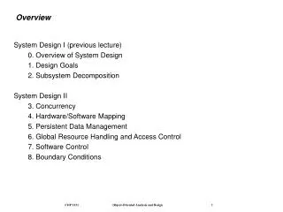
Overview • Neuroanatomy • History • Physical • Clinical Scenarios
Introduction • Facilitates Communication • Provides Baseline • Directs Testing • Identifies Need For Life-Saving Therapies • Risk Management
Risk Management: Case #1 • A 46-year-old female with a long history of migraine headaches presented c/o a severe occipital HA that was different form her past headaches in location and intensity. Neuro exam “WNL”. Patient was treated with Compazine, 10 MG IV, with “Resolution of Headache” and discharged home to “Follow-Up With PMD”. • 18 hours later, patient was brought in by EMS comatose
Risk Management: Case #2 • A 64-year-old male presented with lower back pain which had become progressively worse over the past 2 weeks. The pain was primarily in the lower back without radiation, with nonspecific numbness in the legs. PMH: presently being treated for prostatitis. Exam: “Mild Paralumbar Tenderness”, “SLR -”, “Motor / Sensory Intact”, Knee DTR +2. patient was prescribed Motrin and told to follow-up with his PMD. • Patient developed irreversible renal damage.
Neuroanatomy • Central versus peripheral • symmetrical vs asymmetrical • If central, what is the level: • Cerebrum • Brain Stem • Spinal cord • If peripheral, is it • Nerve • Muscle • NMJ
Central lesions • Lesions in the cerebral cortex result in contralateral deficits of the face and body • Lesions at the midbrain result in contralateral hemiplegia and ipsilateral peripheral paralysis of III and IV • Lesions at the pons result in contralateral hemiplegia and ipsilateral deficits of V, VI, VII, VIII • Lesions at the medulla result in contraleral hemiplegia and ipsilateral deficits of IX, X, XI, XIII
Anatomy of the Spinal Cord • Corticospinal Tracts: motor from cerebral cortex: cross in the lower medulla • Spinothalamic Tracts: pain and temperature: cross 1 or 2 levels above entry • Posterior Column: proprioception and vibration
Spinal Cord : Vascular Supply • Single Anterior • Paired posterior from vertebral arteries (Except in cervical cord) • Radicular Arteries from aorta: • Varying degrees of contribution • Great radicular artery of Adamkiewicz T-10 to L-2 (Major source of blood flow to 50% of anterior cord in 50% of patients) • Anterior perfuses anterior and central cord
UMN vs LMN • UMN increased DTR (after SS) LMN decreased DTR • UMN muscle tone increased LMN tone decreased, atrophy • UMN no fasciculations LMN fasciculations
UMN vs LMN Weakness • Mylopathy = Spinal Cord Process = UMN findings (spasticity, weakness, atrophy, sensory findings, bowel and bladder complaints) • Radiculopathy = Nerve Root Process = LMN findings (Paresthesias, Fasciculations, Weakness, decreased DTR) • Patient may have a radiculopathy with mylopathy below the lesion
The Neuro Exam: History • Neuro complaints may be primary or secondary to other system disease • Infection • Overdose • Metabolic Disorder
The Neuro Exam: History • History often provides the key since the neuro exam may be normal • Subarachnoid Hemorrhage • Carbon Monoxide Poisoning • Subdural Hematoma • Nonconvulsive Seizures
The Neuro Exam: History • Time of Onset • Type of Onset • Progression • Trauma • Associated Symptoms
The Neuro Exam: History • Factors that make it better/worse • Past Symptoms / Events • Past Medical History • Occupational / Environ Exposures
The Neuro Exam: Physical • Vital Signs • Head: Evidence of Trauma • Neck: Bruits, Rigidity • Heart: Murmurs • Abdomen: Masses / Distention • Skin / Scalp: Lesions / Tenderness
The Neuro Exam: Physical • Mental Status • Cranial Nerves • Motor • Sensory • Coordination • Reflexes
The Neuro Exam: Initial Approach • Posture • Decorticate • Decerebrate • Facial or body asymmetry • Hemiparesis results in external rotation of the foot of the affected side
Mental Status Exam • AVPU • GCS • Orientation • Speech (dysarthria vs aphasia) • Comprehension
Mental Status Exam • Confusion assessment method (CAM) • Acute onset / fluctuating course • Inattention • Disorganized thinking • Altered level of consciousness • Mini-mental status exam • Score affected by education and age • <20 = cognitive impairment
Acute Altered Mental Status • Intracranial lesion • Metabolic disorder • Toxin • Infection • Ictal state • Postictal state • Psychogenic
Cranial Nerve Exam • Focus exam on II - VIII • Symmetrical vs asymmetrical
Evaluation of II, III, IV, VI • Visual acuity • Visual fields • Examine the cornea, pupil, fundi • Check afferent function • Extraocular movements • Accentuated when looking in the direction of the paralyzed muscle • Differentiation can be facilitated by placing a colored glass over one eye
Cranial Nerve II • Visual acuity • Visual fields • Fundoscopy • Swinging flashlight test
III Nerve • Emerges from brainstem next to posterior cerebral artery • May be compressed by herniation • Runs in the lateral wall of the cavernous sinus
III Cranial Nerve • Parasympathetics • Levator Palpebrae • Inferior Obliques, Medial, Inferior, and Superior Rectus Muscles SR IO IO SR LR MR MR LR IR SO SO IR
III Cranial Nerve Paralysis • Ptosis • Dilated Pupil • Paralyzed eye is deviated out and down; SO and LR control eye SR IO IO SR LR MR MR LR IR SO SO IR
III Cranial Nerve Lesions • Progressive lesions after passage through the dura usually usually causes a ptosis and pupil dilatation first • Lesions in the nucleus cause motor deficits first • Intact pupil indicates a peripheral ischemic lesion
IV Cranial Nerve • Superior oblique • Causes eye to turn in and down • When paralyzed, eye can not turn down when it is rotated in SR IO IO SR LR MR MR LR IR SO SO IR
VI Cranial Nerve • Lateral rectus • Long course; goes through the CS, not within the wall • Paralysis impairs abduction SR IO IO SR LR MR MR LR IR SO SO IR
Conjugate Gaze • Controlled by supranuclear connections • Medial longitudinal fasciculus is responsible for coordinating the oculomotor nerves; lesions result in impairment of LR and MR moving in sync, ie, contralateral eye does not pass the midline • Multiple sclerosis
Causes of III, VI, VI CN Paralysis • Isolated cases usually due to vascular causes: HTN, DM, Atherosclerosis • Tumors • Increased intracranial pressure • Colloid cyst of the III ventricle • Wernicke-Korsakoff syndrome • Myasthenia, Botulism • Toxic drug reactions
Cranial Nerve V • Sensory: corneal reflexes • Motor: jaw strength and muscle bulk • Corneal reflex may be abnormal in cerebellopontine angle lesions: test in patients with hearing deficits or vertigo
Cranial Nerve VII • Motor • Smile • Bury eyelashes • Nasolabial fold • Forehead has bihemispheric innervation centrally • Taste anterior 2/3
Cranial Nerves VIII - XII • VIII - vestibular function / hearing • IX - taste / sensation posterior pharynx • X - SCM; chin to the opposite side • XII - tongue
Motor Exam • Strength • Primary concern: can patient breathe • Key test: drift of extremity • Tone • Hypertonia: subacute or chronic corticospinal lesion • Hypotonia: LMN lesion or acute UMN • Rigidity: basal ganglia disease
Motor Exam • Bulk • Wasting correlates with LMN • Fasciculation • Anterior horn cell lesion • Tenderness • Metabolic / inflammatory muscle disease
Motor Exam • 0 = no movement • 1 = flicker but no movement • 2 = movement but can not resist gravity • 3 = movement against gravity but can not resist examiner • 4 = resists examiner but weak • 5 = normal
Sensory Exam • Pain / Temp - cross at entrance, ascend in spinal thalamic tract • Light touch - ascend in posterior column, cross in the brain stem • Vibration - posterior column, cross in the brain stem
Sensory Exam • Dermatomal deficit accompanied with pain suggests peripheral lesion • Central deficits are not dermatomal and usually result in loss of sensation not pain • Thalamic pain syndrome
Sensory Exam • Distribution • Right vs left vs bilateral • Dermatomal • Distal versus proximal • Stocking glove • Cape like • Pinprick versus light touch
Sensory Exam • Double simultaneous testing • Establish sharp / dull • Check cheek, dorsum of hands, dorsum of feet • Test both sides simultaneously with pin • lateralizes pain, significant sensory deficit • initially no lateralization but on repeat 15 sec later, lateralization suggests subtle deficit
Coordination • Requires integration of cerebellar, motor, and sensory functions • Balance requires (2 of 3) • vision • vestibular sense • proprioception • Falling with eyes open or closed = cerebellar • Falling only with eyes closed = posterior column or vestibular
Reflexes • Symmetry / upper vs lower • 0 = absent • 1 = hyporeflexia • 2 = normal • 3 = hyperreflexia • 4 = clonus (usually indicates organic disease) • Superficial reflexes (corneal, pharyngeal, pharyngeal, abdominal, anal, cremasteric, bulbocavernosus) • Pathologic reflexes: babinski
Hysteria (conversion vs malingering) • Blindness: opticokinetic test • Hand drop on face test for coma or UE weakness • Hemianesthesia: if real, patient cannot perform finger-to nose with eyes closed; vibration remains intact (if bony skeleton intact) • Weakness: elbow extension or flexor test; wrist extensor test • Unilateral LE weakness: thigh abduction test, hoover test
Pitfalls In The Neurologic Exam • Not getting a complete history utilizing family or observers • Not performing a systematic exam • Jumping to conclusions before gathering all the data • Misinterpreting old lesions for new • Misinterpreting limitations from pain as neurologic deficits
Pearls • Lesions of the cerebral cortex result in sensory and motor defects confined to the contralateral side of the body • Brain stem and spinal cord lesions result in ipsilateral as well as contralateral defects due to varying patterns of crossover
Pearls • Unilateral pain syndromes without motor deficits suggest possible thalamic pathology • A careful exam of CN II, III, IV, and IV is indicated in patients with headache or suspected processes that cause increased ICP • Testing for pronator drift is the best screen for muscle weakness of central origin