Download
1 / 27
340 likes | 1.17k Views
Introduction. Incidence: 1 in every 30,000 ER visitsLaryngeal injuries in 30 to 70 % in penetrating neck trauma (especially zone II)Blunt and penetrating neck injuryAirwayMajor vascular structuresCervical esophagusCervical spine. . Laryngeal Embryology. 3rd and 5th branchial arches 3rd weekRespiratory primordium is derived from primitive foregut4th -5th weeksTracheoesophageal (TE) septum forms by fusion of (TE) folds.

E N D
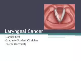
1. Laryngeal Trauma Jean Paul Font, MD
Francis B. Quinn, Jr., MD
Grand Rounds Presentation
Department of Otolaryngology
University of Texas Medical Branch at Galveston
March 28, 2007
2. Introduction Incidence: 1 in every 30,000 ER visits
Laryngeal injuries in 30 to 70 % in penetrating neck trauma (especially zone II)
Blunt and penetrating neck injury
Airway
Major vascular structures
Cervical esophagus
Cervical spine.
3. Laryngeal Embryology 3rd and 5th branchial arches
3rd week
Respiratory primordium is derived from primitive foregut
4th -5th weeks
Tracheoesophageal (TE) septum forms by fusion of (TE) folds
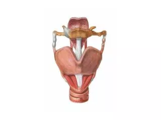
4. Anatomy Support: Hyoid, thyroid, cricoid
Protection of the larynx
Superiorly by the mandible
Inferiorly by the sternum
Laterally by the sternomastoid muscle
Posteriorly by the cervical spine
Innervation: RLN, SLN
5. Anatomy Supraglottis
External support
Soft tissue attachments
Glottis
Relies on external support
Narrowest in the adult
Susceptible to obstruction
Subglottis
Cricoid-narrowest in infants
6. Laryngeal Function Function
Breathing passage
Airway protection
Clearance of secretions
Vocalization
7. Mechanism of Injury Blunt trauma
MVA
Clothesline
Crushing
Strangulation injuries
Penetrating trauma
GSW- related to the type of weapon
Directly penetration or indirectly by the blast effect
Knives Eddie Griffin destroyed a Ferrari Enzo worth $1.5 million Eddie Griffin destroyed a Ferrari Enzo worth $1.5 million
9. Mechanism of Injury Blunt injuries
Most commonly from motor vehicle accidents
Forward thrust
Neck extension impacting steering wheel
Removes the mandibular barrier
Laryngeal skeleton is compressed between a foreign object (i.e., steering wheel or dashboard) and the anterior aspect of the cervical spine
Decrease incidence- seat belt harness and air bags
10. Initial Evaluation ATLS principles
Intubation hazardous
Schaefer in 1991- worsen preexisting injury
Further tears or cricotracheal separation
Respiratory distress
Tracheotomy under local anesthesia
Avoid cricothyroidotomies
Worsen injury
If no acute breathing difficulties
Detailed history and careful physical examination
11. Pediatric patient Blunt pediatric neck injuries
Uncommon the larynx lies higher than the adult
Protected by the mandible
More often life-threatening
Significant injury including laryngotracheal disruption
Smaller cross-sectional area of the pediatric population
Rigid bronchoscopy followed by tracheotomy over the bronchoscope
12. Diagnosis History
Change in voice
Pain
Dyspnea
Dysphagia
Odynophagia
Hemoptysis
Inability to tolerate the supine position Physical Exam
Respiratory rate (saturations)
Stridor
Neck skin
Contusions, Abrasions or Line pattern
Subcutaneous emphysema
Tracheal deviation
Open wound
Air bubbles
Exposed tracheal cartilage
Don�t probe open wounds
May dislodge a hematoma
13. Diagnosis Unstable
Tracheotomy and neck exploration
Stable patients
Flexible fiberoptic laryngoscopy in the ER
CT scan, direct laryngoscopy, bronchoscopy and esophagosopy
14. Ct Scan CT allows:
Evaluation of the laryngeal skeletal framework
Noninvasive avoiding unnecessary operative explorations
15. CT Scan Reserved
Suspected laryngeal injury by history and physical examination
No obvious surgical indications
16. Laryngotracheal Injury Classification Group I injuries
No fracture
Minor hematoma, edema or laceration
Group II injuries
Nondisplaced fractures
Edema or hematoma
Minor mucosal disruption without exposed cartilage
Group III injuries
Displaced fractures
Massive edema or mucosal disruption
Exposed cartilage and/or cord immobility
Group IV injury (group III)
Addition of two or more fracture lines
Skeletal instability or significant anterior commissure trauma
Complete laryngotracheal separation
18. Medical Management Group I injuries
Minimum of 24 hours of close observation
Head of bed elevation
Voice rest
Humidified air
Anti-reflux medication
Serial flexible fiberoptic exams Antibiotics for laryngeal mucosa disruption
19. Steroid Controversial
Early systemic steroids therapy are often given to reduce laryngeal edema
One randomized controlled trial (Ghorayeb 1985)
Intravenous dexamethasone for preventing traumatic laryngeal edema in pediatric bronchoscopy
This study showed no reduction in postbronchoscopy laryngeal edema with the use of intravenous dexamethasone
20. Surgical Management Hemostasis
Evacuation of hematoma
Reconstruction of the laryngeal framework
Coverage of de-epithelialized surfaces
Group II to V required surgical intervention
Surgical options
Endoscopy alone
Endoscopy with exploration
Endoscopy with exploration and stenting
21. Surgical Management Any doubt about the extent of injury endoscopy should be performed
Indications for surgical exploration include:
Large mucosal lacerations
Exposed cartilage
Multiple or displaced cartilaginous fractures
Vocal cord immobility
Fractured cricoid
Disruption of the cricoarytenoid joint
Lacerations involving the free margin of the vocal cord or anterior commisure
Explore within 24 hours of the injury
Maximize airway and phonation results
22. Surgical Management Laryngeal skeleton is exposed from the hyoid to sternal notch
Midline thyrotomy
May use a vertical fracture (2 to 3mm of midline)
Nondisplaced fractures
Suture outer perichondrium
Primary closure with nonabsorbable sutures
Debridement should be minimized
Mucosal lacerations
Meticulously repaired using fine absorbable sutures
Knots outside the laryngeal lumen (prevent granulation)
23. Surgical Management Displace fractures of the cartilages are reduced
Stabilized using stainless steel wires, nonabsorbable suture or miniplates.
Small fragments of cartilage with no intact perichondrium are removed to prevent chondritis.
Anterior commissure- suspend the anterior true vocal cords to the outer perichondrium of the thyroid cartilage
Close the thyrotomy
Nonabsorbable suture, wires or miniplates
24. Surgical Management Endolaryngeal stenting
Disruption of the anterior commissure
Massive mucosal injuries
Comminuted fractures of the laryngeal skeleton
From the false vocal fold to the first tracheal ring
Stability and prevent endolaryngeal adhesions
Removed in a period of 10 to 14 days to prevent mucosal damage
25. Stents Types of stents
Endotracheal tube (COVER THE TOP END TO PREVENT ASPIRATION)
Finger cots filled with gauze or foam
Polymeric silicone stents
Secure the stent
Heavy, nonabsorbable suture
Larynx at the ventricle
Cricothyroid membrane
Tied outside the skin
Endoscopically removed
26. Conclusion Laryngeal trauma although uncommon can be life-threatening
Recognizing any airway compromise and need for immediate intervention could prevent immediate death as well as acute and long term morbidity
Initial management should follow ATLS principles
Most authors agree that tracheotomy should be performed on patients exhibiting respiratory distress
In patients with no acute breathing difficulties, a detailed history, careful physical examination and appropriate diagnostic tools should be use to differentiate the need for medical from surgical management
28. References Schaefer, S.D. Use of CT Scanning in the management of the acutely injured larynx. Otolaryng Clinics NA. Vol 24(1): 31-36. February 1991.
Perdiki, G. Blunt Laryngeal Fracture: Another Airbag Injury The Journal of Trauma: Injury, Infection, and Critical Care. Vol. 48, No. 3. p544-546. 2000
Hwang, S. Y. Management dilemmas in laryngeal trauma
The Journal of Laryngology & Otology., Vol. 118, pp. 325�328. May 2004
Verschueren,D. S. Management of Laryngo-Tracheal Injuries Associated With
Craniomaxillofacial Trauma. American Association of Oral and Maxillofacial Surgeons. P203-214. 2006
Ford, H. Laryngotracheal Disruption From Blunt Pediatric Neck Injuries: Impact of Early Recognition and Intervention on Outcome. Journal of Pediatric Surgery, Vo130, No 2: pp 331-335. (February), 1995
Goudy, S. L. Neck Crepitance: Evaluation and Management of Suspected Upper
Aerodigestive Tract Injury. Laryngoscope 112. p791-795: May 2002
O�Mara, W and Hebert, F. External laryngeal trauma. J La State Med Soc. Vol 152(5): 218-222. May 2000.
Schaefer, S.D. The treatment of acute external laryngeal injuries. Arch Otolaryng HNS. Vol 117: 35-39. January 1991
Cummings: laryngeal Injury. Otolaryngology: Head & Neck Surgery, 4th ed. Mosby, Inc, 2005. 4223-4238
Fuhrman, G.M., Stieg, F.H., and Buerk, C.A. Blunt laryngeal trauma: Classification and management protocol. J Trauma. Vol 30(1): 87-92. January 1990
Ghorayeb BY, Shikhani AH. The use of dexamethasone inpediatric bronchoscopy. J Laryngol Otol 1985;99:1127�9