Download
1 / 1

E N D
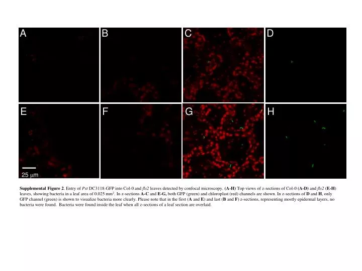
25 m A B C D E F G H Supplemental Figure 2. Entry of Pst DC3118-GFP into Col-0 and fls2 leaves detected by confocal microscopy. (A-H) Top views of z-sections of Col-0 (A-D) and fls2 (E-H) leaves, showing bacteria in a leaf area of 0.025 mm2. In z-sections A-C and E-G, both GFP (green) and chloroplast (red) channels are shown. In z-sections of D and H, only GFP channel (green) is shown to visualize bacteria more clearly. Please note that in the first (A and E) and last (B and F) z-sections, representing mostly epidermal layers, no bacteria were found. Bacteria were found inside the leaf when all z-sections of a leaf section are overlaid.