Download
1 / 24
1.31k likes | 4.73k Views
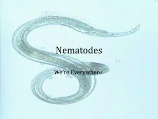
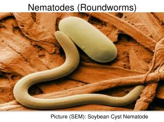
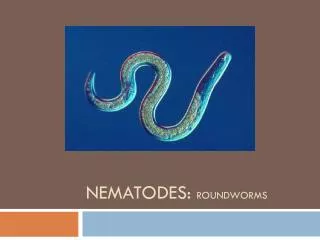
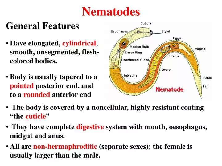
Nematodes. General Features Have elongated, cylindrical , smooth, unsegmented , flesh-colored bodies. Body is usually tapered to a pointed posterior end, and to a rounded anterior end. The body is covered by a noncellular , highly resistant coating “the cuticle ”

E N D
Nematodes General Features • Have elongated, cylindrical, smooth, unsegmented, flesh-colored bodies. • Body is usually tapered to a pointed posterior end, and to a rounded anterior end • The body is covered by a noncellular, highly resistant coating “the cuticle” • They have complete digestive system with mouth, oesophagus, midgutand anus. • All are non-hermaphroditic (separate sexes); the female is usually larger than the male.
Nematodes General Features • Infection occurs either by eating uncooked meat with larvae in it, by entrance directly through the skin or cuts, or by transfer via blood-sucking insects • They are classified into 2 main categories according to their primary location: • Intestinal nematodes • Tissue nematodes (filariae)
A. Intestinal Nematodes Large size, cylindrical Most adult worms live in the intestinal tract Most of their diseases are diagnosed by identifying their characteristic eggs in stool B. Tissue Nematodes Elongated, cylindrical Inhabit either lymph vessels; or skin and subcutaneous tissues Diseases are diagnosed by demonstrating microfilariae in blood, in tissue or tissue fluids Main features of Nematodes
Medically important nematodes. • Intestinal Nematodes • 1. Enterobius vermicularis • 2. Ascaris lumbricoides • 3. Strongyloides stercolaris • 4. Hookworms • *Ancylostoma duodenal • * Necator americanus • 5. Trichuris trichiuria • 6. Trichinella spiralis B. Tissue Nematodes Filarial worms are classified according to their habitat into: I.Lymphatic filariae: 1. Wuchereriabancrofti 2. Burgiamalayi II. Cutaneousfilariae: 1. Dracunculusmedinensis 2. Loa loa 3. Onchocercavolvulus
Type A: Intestinal Nematodes Life cycle of intestinal nematodes type A. • The worm develops in intestine • Eggs are released with feces into environment • Mature eggs are ingested by new host and hatch in intestine giving larvae
Enterobius vermicularis Female Male Female Male Female E. vermicularis Ascaris lumbricoides Ascaris fertilized Egg E. vermicularis Egg
Ascaris lumbricoides Enterobius vermicularis Ascariasis, roundworm ascariasis Embryonated egg Ingestion of eggs in food contaminated with human feces Small intestine, lung Egg in feces; are oval, brown with colorless shell containing very large fertilized unsegmented ovum • Pinworm, oxyuris, seatworm • enterobiasis (oxyuriasis) • Embryonated egg • Ingestion; or autoinfection via nails scratching the perianus • Large intestine • Egg in nails or perianus area. Eggs are colorless, asymmetric with one flattened side
migrating worms cause occlusion of biliary tract or oral expulsion in lung it causes inflammation with pulmonary symptoms, e.g. cough, hemoptysis Characteristic eggs in feces, larvae identified in sputum or gastric aspirate adult worm may pass in stool. Albendazole, mebendazole, pyrantel pamoate • perianal pruritis, especially at night, appendicitis, abdominal pain, *invasion of girls’ genital tract causes vaginitis, pelvic or peritoneal granulomas • Characteristic eggs collected in mornings from perianal area using transparent adhesive tape adult worm may be found in perianal area or during vaginal examination • Pyrantel pamoate
Type B: Intestinal Nematodes Life cycle of intestinal nematodes type B. • The worm develops in intestine • Eggs are released with feces into environment • Larvae hatch and develop in environment • Infection occurs through skin penetration by larvae
Hookworms Ancylostomaduodenale Old World hookwormNecator americanusNew World hookworm
Disease • Infective stage • Mode of transmission • Site of infection • Diagnostic stage • Clinical findings • Laboratory diagnosis • Treatment Hookworm infection Filariform larva Filariform larvae in moist soil penetrate skin through bare feet Small intestine, heart, lung Egg in feces Iron deficiency anemia due to loss of blood at site of attachment in intestine *cardiac problems *local skin manifestations “ground itch” *respiratory symptoms during larval pulmonary attack Characteristic egg in stool Albendazole, mebendazole, pyrantel pamoate
Common name • Disease • Infective stage • Mode of transmission • Site of infection • Diagnostic stage • Clinical findings • Laboratory diagnosis • Treatment Trichinosis Trichinillosis, trichinosis Encysted larva Larvae in undercooked pork. Pigs are main reservoir Striated muscles & small intestine Larvae in muscles and tissues Larvae migration in muscular tissues cause facial and periorbitaloedema, rash, muscle pain, conjunctivitis *muscle biopsy to identify larvae in striated muscles *indication of eosinophilia *serologic tests Steroids plus mebendazole in severe infections *Thiabendazole
Tissue (Filarial) Nematodes Lymphatic Wuchereria bancrofti Cutaneous Dracunculus medinensis Loa loa Onchocerca volvulus
Tissue (Filarial) Nematodes Lymphatic Wuchereria bancrofti
Wuchereria bancrofti • Distribution: worldwide in tropical areas • Disease: Elephantiasis, WuchereriasisBancroftianfilariasis, lymphatic filariasis • Vector: mosquito (Anopheles sp. or Culexsp.) • Infective stage: L3 larvae • Diagnostic stage: motile microfilaria • Habitat: lymph nodes, lymphatic glands and vessels in legs, arms and genitalia (testes) • Symptoms: Inflammation of vessels, rupture of lymphs, fibrosis, leading to obstruction. Thickening, hypertrophy of tissues, enlargement of tissues (especially extremities and genitalia)
Diagnosis: demonstration of microfilaria in blood molecular diagnosis using PCR Treatment: diethylcarbamazine, surgery in elephantiasis
Tissue (Filarial) Nematodes Cutaneous Loa loa
Common name • Geog. Distrib. • Vector • Site of infection • Disease • Symptoms • Diagnosis • Treatment eye worm Africa fly of genus Chrysops (deerfly or mango fly) subcutaneous tissues, and muscles loiasis, Calabar swelling angioedema, swelling of various parts of body (Calabar swelling), conjunctivitis, irritation, oedema of eyelids, impaired vision demonstration of microfilaria in blood (since adult worm lives in subcutaneous tissue and microfilaria in blood) diethylcarbamazine for microfilaria and adult, soothing lotion for Calabar swelling