Download
1 / 48
480 likes | 489 Views
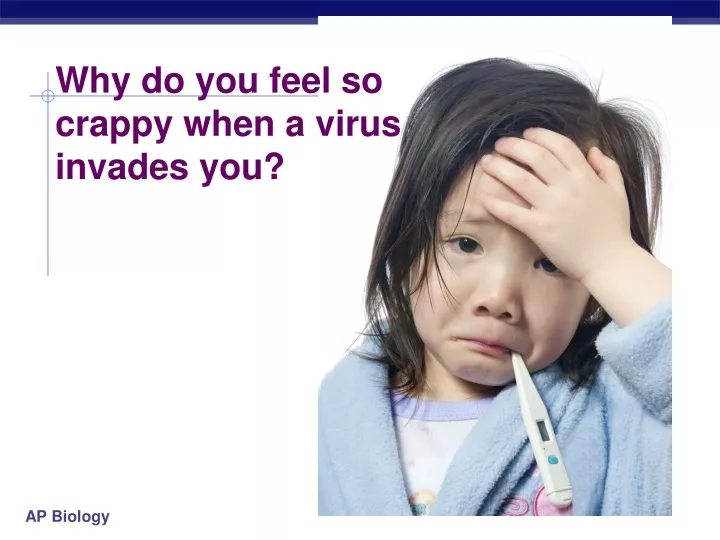
Why do you feel so crappy when a virus invades you?. phagocytic leukocyte. Fighting the Enemy Within !. The Lymphatic System. lymphocytes attacking cancer cell. lymph system. The Lymphatic System.

E N D
phagocytic leukocyte Fighting theEnemy Within! The LymphaticSystem lymphocytes attacking cancer cell lymph system
The Lymphatic System • a series of glands and intercellular fluid that creates and maintains the cells of the immune system • Lymph = a clear fluid that circulates among your cells outside of the circulator system (like water moving in a sponge)
Disease: Any disruption in homeostasis • Can be communicable (contagious, e.g. the common cold) or non-communicable (e.g. cancer). • The immune and lymphatic systems combat disease and attempt to restore homeostasis.
Germ Theory: Communicable disease is caused by microscopic organisms called pathogens • Viruses – non-living infectious particles; invade the cell itself; antibiotics are ineffective • Bacteria – prokaryotic cell species that invade the body only; antibiotics are effective as long as the bacteria is not resistant
Virus Types • Lytic – invade cell and kill instantly to create new viruses (e.g. the flu) • Lysogenic – invade cell and enter the nucleus to become part of the genome; eventually turn lytic, but may lie dormant for years (e.g. HIV)
Avenues of attack • Points of entry • digestive system • respiratory system • urogenital tract • break in skin • Spreading through the body • circulatory system • lymph system
Immune System • Protect against attack from outside • Defend the body against pathogen invaders like… • viruses • HIV, flu, cold, measles, chicken pox • bacteria • pneumonia, meningitis, tuberculosisLyme disease • fungi • yeast (“Athlete’s foot”…) • protists • amoeba, malaria • And attack from inside • cancers = abnormal body cells
Lines of defense • 1st line: Non-specific barriers • Barriers against entry; skin and mucus layers • 2nd line: Non-specific patrols • Cells that monitor the circulatory systems looking for ANYTHING that doesn’t belong; called leukocytes (phagocytic white blood cells) • 3rd line: Specific patrols, true immunity • Lymphocytes & antibodies; cells that remember pathogens they’ve encountered before and attack faster than the WBC
Immune system must be able to distinguish between self and non-self and recognize new pathogens • Innate immunity – the nonspecific WBCs that all individuals are born with • Acquired immunity – as the body is exposed to pathogens, lymphocytes antibodies accumulate and strengthen immunity; why vaccines work
Red blood cells and all the different types of White blood cells come from stem cells inflammatory response Red blood cells fightparasites Lymphocytes short-lived phagocytes 60-70% WBC develop into macrophages
The lymphatic system (lymph nodes) controls and monitors the production of your WBC Interstitialfluid Bloodcapillary Adenoid Tonsils Lymphaticvessels Thymus Lymphatic vessel Tissuecells Lymphatic vessel Peyer’spatches(smallintestine) Spleen Lymphnodes Appendix(cecum) Lymphnode Masses ofdefensive cells
Examples: Non-specific External defense Lining of trachea: ciliated cells & mucus secreting cells • Barrier • skin • Traps • mucous membranes, cilia,hair, earwax • Elimination • coughing, sneezing, urination, diarrhea • Unfavorable pH • stomach acid, sweat, saliva, urine • Lysozyme enzyme • digests bacterial cell walls • tears, sweat
Examples: Non-specific patrolling cells bacteria • Patrolling cells & proteins • attack pathogens, but don’t “remember” for next time • leukocytes • phagocytic white blood cells • macrophages, neutrophils, natural killer cells • complement system • Free-floating proteins that destroy cells • inflammatory response • increase in body temp. • increase circulation • attract macrophages (WBC) macrophage yeast
Leukocytes: Phagocytic WBCs • Attracted by chemical signals released by damaged cells • Neutrophils • most abundant WBC (~70%) • ~ 3 day lifespan • phagocyte • Macrophages • “big eater” phagocyte • long-lived • Natural Killer Cells • destroy virus-infected cells & cancer cells
Destroying self cells gone bad! • Natural Killer Cells perforate cells • release perforin protein • insert into membrane of target cell • forms pore allowing fluid to flow in & out of cell • cell ruptures (lysis) • apoptosis vesicle natural killer cell perforin cell membrane perforinpuncturescell membrane cell membrane virus-infected cell
Anti-microbial proteins • Complement system • ~20 proteins circulating in blood plasma • attack bacterial & fungal cells • form a membrane attack complex • perforate target cell • apoptosis • cell lysis extracellular fluid complement proteinsform cellular lesion plasma membrane of invading microbe complement proteins bacterial cell
Inflammatory response • Damage to tissue triggers local non-specific inflammatory response • release chemical signals • histamines & prostaglandins • capillaries dilate, becomemore permeable (leaky) • delivers macrophages, RBCs, platelets, clotting factors • fight pathogens • clot formation • increases temperature • decrease bacterial growth • stimulates phagocytosis • speeds up repair of tissues
Figure 43.8-1 Pathogen Splinter Macro-phage Signalingmolecules Mastcell Capillary Neutrophil Redblood cells
Figure 43.8-2 Pathogen Splinter Movementof fluid Macro-phage Signalingmolecules Mastcell Capillary Neutrophil Redblood cells
Figure 43.8-3 Pathogen Splinter Movementof fluid Macro-phage Signalingmolecules Mastcell Phagocytosis Capillary Neutrophil Redblood cells
Fever • When a local response is not enough • system-wide response to infection • activated macrophages release interleukin-1 • triggers hypothalamus in brain to readjust body thermostat to raise body temperature • higher temperature helps defense • inhibits bacterial growth • stimulates phagocytosis • speeds up repair of tissues • causes liver & spleen to store iron, reducing blood iron levels • bacteria need large amounts of iron to grow
Example: Acquired Immunity • Specific defense with memory • lymphocytes • B cells • antibodies(a.k.a. immunoglobulins) • T cells • Responds to… • antigens • cellular name tags • specific pathogens • specific toxins • abnormal body cells (cancer) B cell
How are invaders recognized? • Antigens • cellular name tag proteins • “self” antigens • no response from WBCs • “foreign” antigens • response from WBCs • Used to mark pathogens…: viruses, bacteria, protozoa, parasitic worms, fungi, toxins • Or non-pathogens: cancer cells, transplanted tissue, pollen “self” “foreign”
bone marrow Lymphocytes • B cells • mature in bone marrow • humoral response system • “humors” = body fluids • attack pathogens still circulating in blood & lymph • produce antibodies • T cells • mature in thymus • cellular response system • attack invaded cells • “Maturation” • learn to distinguish “self” from “non-self” antigens • if react to “self” antigens, cells are destroyed during maturation
Humoral (antibody-mediated) immune response Cell-mediated immune response Key Figure 43.20 Antigen (1st exposure) Stimulates Engulfed by Gives rise to Antigen-presenting cell Helper T cell Cytotoxic T cell B cell Memoryhelper T cells Antigen (2nd exposure) Memorycytotoxic T cells Active cytotoxic T cells Plasma cells Memory B cells Secretedantibodies Defend against extracellularpathogens Defend against intracellularpathogens and cancer
B cells • Attack, learn & remember pathogens circulating in blood & lymph • Produce specific antibodiesagainst specific antigen • Types of B cells • plasma cells • immediate production of antibodies • rapid response, short term release • memory cells • continued circulation in body • long term immunity
Y Y Y Y Y Y Y Y Y Y Y Y Y Y Y Y Y Y Y Y Y Y Y Y Y Y Y Y Y Y Y Y Y Y Y Y Y Y Y Y Y Y Y Y Y Y Y Y Y Y Y Y Y Y Y Y Y Y Y Y Y Y Y Y Y Y Y Y Y Y Y Y Y Y Y Y Y Y Y Y Y Y Y Y Y Y Y Y Y Y Y Y Y Y Y Y Y Y Y Y Y Y Y Y Y Y Y Y Y Y Y Y Y Y Y Y Y Y Y Y Y Y Y Y Y Y Y Y Y Y Y Y Y Y Y Y invader(foreign antigen) B cells + antibodies memory cells “reserves” recognition Y capturedinvaders Y Y Y Y Y Y Y Y Y Y Y Y Y Y Y Y Y macrophage Y Y Y Y Y Y Y Y Y Y clones 1000s of clone cells plasma cells release antibodies 10 to 17 days for full response B cell immune response tested by B cells (in blood & lymph)
Y Y Y Y Y Y Y Y Y Y Y Y Y Y Y Y Y Y Y Y Y Y Y Y Y Y Y Y Y Y Y Y Y Y Y Y Antibodies Y Y • Proteins that bind to a specific antigen • multi-chain proteins • binding region matches molecular shape of antigens • each antibody is unique & specific • millions of antibodies respond to millions of foreign antigens • tagging “handcuffs” • “this is foreign…gotcha!” Y Y Y antigen-binding site on antibody Y antigen Y Y Y variable binding region Y Y each B cell has ~50,000 antibodies
Y Y Y Y Y Y Y Y Y Y Y Y Y Y Y Y antigen-binding site variable region s s s s s s s s light chain s s s s s s s s s s s s s s s s heavy chains s s s s light chains s s s s s s s s antigen-binding site antigen-binding site heavy chains Structure of antibodies Y Y Y Y Y Y Y Y Y light chain B cell membrane
Y Y Y Y Y Y invading pathogens tagged with antibodies macrophageeating tagged invaders What do antibodies do to invaders? neutralize capture precipitate apoptosis
Exposure to antigen IgM IgG Antibody levels 0 2 4 6 Weeks Y Y Y Y Y Y invading pathogens tagged with antibodies Classes of antibodies macrophageeating tagged invaders • Immunoglobulins • IgM • 1st immune response • activate complement proteins • IgG • 2nd response, major antibody circulating in plasma • promote phagocytosis by macrophages • IgA • in external secretions, sweat & mother’s milk • IgE • promote release of histamine & lots of bodily fluids • evolved as reaction to parasites • triggers allergic reaction • IgD • receptors of B cells???
Figure 43.UN02 Stem cell Cell division andgene rearrangement Elimination ofself-reactiveB cells Antigen Clonalselection Formation ofactivated cellpopulations Antibody Plasma cells Memory B cells Pathogen Receptors bind to antigens
Vaccinations • Immune system exposed to harmless version of pathogen • stimulates B cell system to produce antibodies to pathogen • “active immunity” • rapid response on future exposure • creates immunity without getting disease! • Most successful against viruses
Albert Sabin 1962 oral vaccine 1914 – 1995 Jonas Salk April 12, 1955 • Developed first vaccine • against polio • A disease that attacks motor neurons
Passive immunity • Obtaining antibodies from another individual • maternal immunity • antibodies pass from mother to baby across placenta or in mother’s milk • critical role of breastfeeding in infant health • mother is creating antibodies against pathogens baby is being exposed to • Injection • injection of antibodies • short-term immunity
T What if the attacker gets past the B cells in the blood & actually infects (hides in) some of your cells? You need trained assassins to recognize & kill off these infected cells! Attackof the Killer T cells! But how do T cellsknow someone ishiding in there?
MHC protein T or Bcell MHC proteins displaying self-antigens How is any cell tagged with antigens? • Major histocompatibility (MHC) proteins • proteins which constantly carry bits of cellular material from the cytosol to the cell surface • “snapshot” of what is going on inside cell • give the surface of cells a unique label or “fingerprint” Who goes there? self or foreign?
MHC proteins displaying foreign antigens TH cell WANTED How do T cells know a cell is infected? • Infected cells digest some pathogens • MHC proteins carry pieces to cell surface • foreign antigens now on cell membrane • called Antigen Presenting Cell (APC) • macrophages can also serve as APC • tested by Helper T cells infectedcell T cell with antigen receptors
T cells • Attack, learn & remember pathogens hiding in infected cells • recognize antigen fragments • also defend against “non-self” body cells • cancer & transplant cells • Types of T cells • helper T cells • alerts rest of immune system • killer (cytotoxic) T cells • attack infected body cells • memory T cells • long term immunity T cell attacking cancer cell
Figure 43.12 Displayedantigenfragment T cell T cell antigenreceptor MHC molecule Antigenfragment Pathogen Host cell (a) Antigen recognition by a T cell Top view Antigen fragment MHCmolecule Host cell (b) A closer look at antigen presentation
Attack of the Killer T cells • Destroys infected body cells • binds to target cell • secretes perforin protein • punctures cell membrane of infected cell • apoptosis vesicle Killer T cell Killer T cellbinds toinfected cell cell membrane perforinpuncturescell membrane cell membrane infected cell destroyed target cell
Immune system & Blood type Matching compatible blood groups is critical for blood transfusions A person produces antibodies against foreign blood antigens
Immune system malfunctions • Auto-immune diseases • immune system attacks own molecules & cells • lupus • antibodies against many molecules released by normal breakdown of cells • rheumatoid arthritis • antibodies causing damage to cartilage & bone • HIV • Helper T cells attacked by virus • multiple sclerosis • T cells attack myelin sheath of brain & spinal cord nerves • Allergies • over-reaction to environmental antigens • allergens = proteins on pollen, dust mites, in animal saliva • stimulates release of histamine
Figure 43.22 Histamine IgE Allergen Granule Mast cell
HIV & AIDS – A disease of the Immune System • Human Immunodeficiency Virus • virus infects helper T cells • helper T cells don’t activate rest of immune system: killer T cells & B cells • also destroys helper T cells • AIDS: Acquired ImmunoDeficiency Syndrome • infections by opportunistic diseases • death usually from • “opportunistic” infections • pneumonia, cancers HIV infected T cell
Epidemiology • The study of disease outbreaks to determine cause and cure • Center for Disease Control (CDC) • Develop vaccines, manage outbreaks, advise public health institutions, protect against biowarfare