Download
1 / 13
130 likes | 240 Views
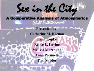
A Comparative Analysis of Centrosome and Soma Migration in Neurons. Rachel Boerner Calvin College Richard Ward Computational Sciences and Engineering Ryan Kerekes Measurement Science and Systems Engineering August 2009. Overview. Background Previous work Methods Results Future work.

E N D
A Comparative Analysis of Centrosome and Soma Migration in Neurons Rachel Boerner Calvin College Richard Ward Computational Sciences and Engineering Ryan Kerekes Measurement Science and Systems Engineering August 2009
Overview • Background • Previous work • Methods • Results • Future work
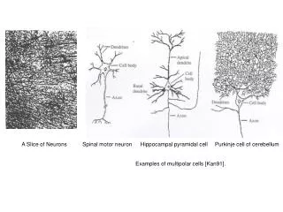
Neuron structure Soma Axon Dendrites • Basic structures • Somas • Axons • Dendrites • Centrosomes http://serendip.brynmawr.edu/bb/kinser/definitions/def-neuron.html Actual images of cerebral neurons
Neuron function and migration • Function determines structure • Migrate Examples of different neuron structures based on their function Migrating neuron along radial glial cell http://glia-uab.infomedia.com/content.asp?id=104968 http://www.mind.ilstu.edu/curriculum/neurons_intro/neurons_intro.php
St. Jude Children’s Research Hospital Neurons migrate during the utero stage or early years of development Abnormal neuron migration has been linked to certain diseases St. Jude’s interested in tracking the neurons for diagnostic purposes Images taken with a Marianas spinning disk confocal microscope Hundreds of images waiting to be manually analyzed Weeks to go through one data set
Oak Ridge National Laboratory • Centrosome detection and tracking • Filter • Threshold • Search for small, round, bright objects • Use joint probabilistic data association filter (above) The right image shows the detections made by Ryan's code. The left image is those detections locations superimposed on the original image. (above) A three dimensional plot of centrosome movement in the x-, y-, and z-directions.
Current Work • Different color schemes show more details
Method • Attempted method • Gradient and fill • Centroid Sequence of attempted code for soma segmentation • Actual method • Euclidean transform equation • Thresholding (left) An image after a rotationally symmetric Gaussian lowpass filter is applied. (center) The image after thresholding for intensity and distance from the center of an object. (right) The segmentation of the somas based on regions being larger than a certain radius.
Results (below) This graph is the plot of the motion of the soma throughout the various timeframes. Each pixel corresponds to 0.15 μm. • Analyzed visually • See random motion • Centrosomes move at an average speed of 0.1 μm • Somas move at an average speed of 0.03 μm • Not much correlation between soma and centrosome migration • Chemicals inhibited cell motion • Centrosome and soma pairs are difficult to match (above) The graphs are 3D images of the detections and tracks of somas and centrosomes. The red marks the soma tracks and the blue corresponds to the centrosome tracks. The images were taken ever 16 seconds.
Conclusion • Developed code for soma segmentation • Detected no correlation between soma and centrosome motion Future Work • Analyze neuron migration in three dimensions • Study and track actin movement • Recognize patterns of behavior between neuron migration and specific diseases • Build techniques for the alteration or prevention of abnormal neuron migration
Bibliography R. A. Kerekes, S. S. Gleason, N. Trivedi, and D. J. Solecki, “Automated 3-D tracking of centrosomes in sequences of confocal image stacks,” submitted to EMBC `09, Minneapolis, 2009. "Neuron." Serendip. 2003. 20 July 2009 <http://images.google.com/imgres?imgurl=http://serendip.brynmawr.edu/bb/kinser/definitions/neuron1.gif&imgrefurl=http://serendip.brynmawr.edu/bb/kinser/definitions/def-neuron.html&usg=__sDy8e5q728xNzj7>. McCoy, Eric. "Glia as Guides for Neuronal Migration,." UAB Center for Glial Biology in Medicine. UAB. 22 July 2009 <http://glia-uab.infomedia.com/content.asp?id=113339>. "ORNL, St. Jude track neurons to predict and prevent disease." The Oak Ridger, 30 March 2009, sec. A3. Stufflebeam, Robert. "Neurons, Synapses, Action Potentials, and Neurotransmission." Consortium on Cognitive Science Instruction. 2008. The Mind Project. 22 July 2009 <http://www.mind.ilstu.edu/curriculum/neurons_intro/neurons_intro.php>.
Acknowledgements I would like to thank Richard Ward and Ryan Kerekes for the opportunity to work on this project. Also, thanks to Debbie McCoy, who made provisions for this research experience.