Download
1 / 64
770 likes | 1.33k Views
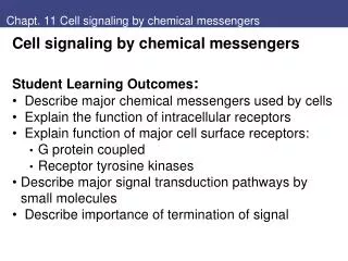
Chapt. 15 Cell Signaling. Student Learning Outcomes: Transmembrane proteins communicate signals from outside the cell to the inside 1*. Explain various signaling molecules and their receptors: how cells receive information from outside.

E N D
Chapt. 15 Cell Signaling • Student Learning Outcomes: • Transmembrane proteins communicate signals • from outside the cell to the inside • 1*. Explain various signaling molecules and their receptors: • how cells receive information from outside. • 2*. Explain several ways signals to receptors are transmitted • through the cell (second messenger pathways) • 3. Describe examples of signal transduction • 4. Describe involvement of cytoskeleton in signal transduction.
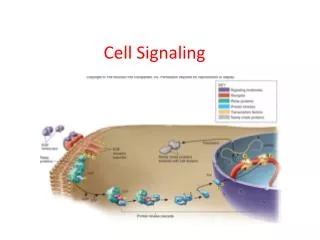
Fig 15.1 Modes of cell-cell signaling 1. Signaling molecules and receptors: Modes of cell signaling include: • Direct cell-cell interaction (ex. Integrins, cadherins) • Secreted molecules: • Endocrine – distant • Estrogen hormone • Paracrine – local • neurotransmitter • Autocrine – self • T cell response • Cancer cells Fig. 15.1
Signaling Molecules and Their Receptors Signal molecules bind receptors: Intracellular receptors bind small hydrophobic signaling molecules that cross plasma membrane Ex. nuclear receptors (steroid hormone) Cell surface receptors bind hydrophilic molecules, peptides, growth factors that don’t cross plasma membrane Ex. insulin receptor, epidermal growth factor, neurotransmitters filopdia
Steroid hormones, thyroid hormone, vitamin D3, retinoic acid Nuclear Receptor superfamily: Intracellular receptors bind hydrophobic hormones • steroid hormones • thyroid hormone, • vitamin D3, • retinoic acid • cortisol Receptors are transcription factors, active after binding hormone Fig. 15.2
Histone tails can be marked with acetylation, methylation, phosphorylation Modifications of histone tails can alter gene regulation – bind coactivators, corepressors Figs. 7.34, 35 Acetylation of lysines (HAT) Deaceylation (HDAC)
Fig 15.3 Glucocorticoid action Some nuclear receptors inactive without hormone: • Glucocorticoid receptor (GR) is bound to Hsp90 chaperone in absence of hormone. • Glucocorticoid binding displaces Hsp90 → • GR binds specific DNA sequences, activates transcription • Note coactivator HAT (histone acetyl transferase) Fig. 15.3
Fig 15.4 Gene regulation by thyroid hormone receptor Some nuclear receptors alter activity after hormone • Absence of hormone, thyroid hormone receptor (TR) • Binds DNA and corepressor complex (HDAC) (histone deacetylase) • represses transcription • Hormone binding → TR binds coactivator (HAT) • activates transcription (see Fig. 7.34) Fig. 15.4
Fig 15.5 Synthesis of nitric oxide Nitric oxide (NO)is a paracrine signaling molecule in nervous, immune, circulatory systems • Alters activity of enzymes (guanylylcyclase makes cGMP) • Synthesized from Arg by nitric oxide synthase (NOS) • Only local effects, extremely unstable, t1/2 of seconds • Ex. Dilation of blood vessels (after neurotransmitter signals) CO is also paracrine Fig. 15.5
Fig 15.6 Structure of some neurotransmitters Neurotransmitterscarry signals between neurons, from neurons to other target cells • Small hydrophilic • released after arrival of action potential at end of neuron. • Diffuse across cleft • Bind receptors on target cell surface: ligand-gated ion channels cell-surface receptors • Coupled to G proteins Fig. 15.6
Signaling Molecules and Their Receptors Peptide signaling molecules: Table 1 diverse sizes (5 – 230 amino acids) and roles Peptide hormones Insulin, glucagon, pituitary gland hormones (growth hormone, follicle-stimulating hormone, prolactin) Neuropeptides & neurohoromone Enkephalins and endorphins act as neurotransmitters at synapses, oxytocin stimulates smooth muscle contraction Polypeptide growth factors Nerve growth factor (NGF) Epidermal growth factor (EGF) stimulates cell proliferation
Signaling Molecules and Their Receptors Eicosanoidsarelipid signaling molecules: Prostaglandins and leukotrienes Autocrine or paracrine pathways Inflammation, smooth-muscle contraction, platelet aggregation Synthesized from arachidonic acid (from phospholipid by PLA2) Aspirin (NSAID) targets cyclooxygenase (COX) (1st step in Prostaglandin syn) Fig. 15.8 COX
Signaling Molecules and Their Receptors Plant hormones: small molecules regulate Gibberellins— stem elongation Auxins— cell elongation Ethylene— fruit ripening Cytokinins— cell division Abscisic acid— onset of dormancy Fig. 15.9
Functions of Cell Surface Receptors 2** Functions of cell-surface receptors: Most ligands for cell-cell signaling bind surface receptors on targets Ligand does not enter cell Binding initiates: chain of intracellular reactions (amplify signal) Alter activity of enzymes Affect ion channels Often change in gene expression * 2 Main types: G protein-coupled, Receptor tyr kinase (RTK) Fig. 8.40 ex. signal cascade
G-coupled Surface Receptors Largest family of cell surface receptors. Signals transmitted via guanine nucleotide-binding proteins (G proteins) in cytoplasm Receptors have 7 transmembraneαhelices G proteins discovered during studies of cyclic AMP (cAMP), second messenger that mediates cellular responses to many hormones 2a. G protein-coupled receptors: Fig. 15.11 G-protein coupled receptor
Fig 15.12 Hormonal activation of adenylyl cyclase Binding of ligands induces conformational change: • Cytosolic domain activates G protein on inner face of plasma membrane. • Activated G protein a subunit dissociates from receptor • Carries signal to intracellular target Ex. Heterotrimeric G protein intermediary in activation of adenylyl cyclase, which synthesizes cAMP *Fig. 15.12 G-protein coupled receptor
Fig 15.13 Regulation of G proteins G proteins have 3 subunits: α, β, and γ. • Called heterotrimeric G proteins to distinguish from small guanine nucleotide-binding proteins, Ras (Rab, Ran) • subunit is regulator: Hormone binding stimulates GTP to bind a (exchange GDP); bg dissociate, (interact different targets) GTP hydrolysis terminates (details later) Fig. 15.13 G-protein coupled receptor (ex. a binds adenylyl cyclase)
Functions of G-protein coupled Receptors Different G proteins connect receptors to distinct targets: Humans have 21 a, 6 b, 12 g Ex. enzyme regulation: epinephrine G protein is Gs,: a subunit stimulates adenylyl cyclase G proteins can also regulate ion channels: Ex. Heart muscle cells have different acetylcholine receptor (G protein-coupled) than nerve and skeletal muscle cells (ligand-gated ion channel (Fig. 13.25): αsubunit of this G protein (Gi) inhibits adenylyl cyclase. The Giβγsubunits open K+ channels in plasma membrane, which slows heart muscle contraction.
Key Experiment 15.1 G Protein-Coupled Receptors and Odor Detection: The odorant receptor protein family Largest family of G protein-coupled receptors is responsible for odor detection. • Odorant receptors on surface of olfactory neurons encoded by multigene family (400 humans, 1000 in dogs, rats). • Odor binds receptor on surface of olfactory neurons; stimulates adenylyl cyclase, increased cAMP opens Na+ channel and nerve impulse Buck, Axel 1991 cloned receptors: White conserved aa Black variable
Functions of Cell Surface Receptors 2b. Receptor protein-tyrosine kinases (RTK): Receptors directly link to intracellular enzyme: Largest family phosphorylates substrates on tyrosine residues Receptors for most polypeptide growth factors: EGF, NGF, PDGF, insulin, and others Ligands binding outside: activate cytosolic kinase domains phosphorylation of receptors and binding intracellular targets propagates signal • Fig. 15.14 RTK receptors Conserved structure
Fig 15.15 Dimerization and autophosphorylation of RTK receptor • Ligand-binding induces receptor dimerization. • Receptor autophosphorylation - 2 polypeptide chains cross-phosphorylate one another • Fig. 15.15 RTK receptors dimerize, autophosphorylate
Fig 15.16 Downstream signaling molecules bind RTK receptors Autophosphorylation has two roles: • Tyr PO4 in catalytic domain increases kinase activity • Tyr PO4 outside catalytic domain → binding sites for proteins that transmit downstream signals • Common motif is the SH2 domain on downstream molecule • Fig. 15.16 RTK receptors bind downstream signal proteins
Complex between an SH2 domain and phosphotyrosine peptide Downstream signaling molecules have domains that bind to specific phosphotyrosine-containing peptides • SH2 domainswere the first characterized (~100aa): • initially recognized in nonreceptorprotein-tyrosine kinases related to Src, (oncogenic protein of Rous sarcoma virus) Other proteins bind via PTB domains (phosphotyrosine-binding). Fig. 15.17: SH2 domain: p-tyr: Blue, 3 aa that bind p-tyr peptide (red = P; yellow backbone); Purple is groove
Fig 15.18 Signaling from cytokine receptors 2c. Cytokine receptor superfamily: • Receptors for cytokines (interleukin-2, erythropoietin) and some polypeptide hormones (growth hormone) • Structure similar to receptor protein-tyrosine kinases, but no catalytic activity cytosolic domains • Work with nonreceptor protein-tyrosine kinases; • Phosphorylated receptor binds downstream molecules via SH2 domains. Fig. 15.18
Functions of Cell Surface Receptors Nonreceptor Kinases associated with cytokine receptors belong to Janus kinase (or JAK) family. Members of JAK family appear to be universally required for signaling from cytokine receptors Fig. 15.40 part; More later
Functions of Cell Surface Receptors Other nonreceptor protein-tyrosine kinases belong to Src family, signal downstream of: cytokine receptors, receptor protein-tyrosine kinases, antigen receptors on B and T lymphocytes, integrins at sites of cell attachment to ECM (matrix) Src protein: Oncogene of RSV virus
Functions of Cell Surface Receptors 2d. Receptors linked to other enzymatic activities: Protein-tyrosine phosphatases: remove phosphate groups from phospho-tyr, counterbalance effects of protein-tyrosine kinases Protein-serine/threonine kinases Ex. Receptors for transforming growth factor β(TGF- β) polypeptide - growth factor controls cell proliferation Receptor guanylylcyclases- cytosolic domain catalyzes formation of cyclic GMP, anothersecond messenger Some receptors have associated protease: Ex. Tumor Necrosis factor (TNF) binding receptor induces cell death (apoptosis; Chapt. 17); downstream proteases.
Pathways of Intracellular Signal Transduction 15.3 Intracellular signal transduction: Chain of reactions transmits signals from cell surface to intracellular targets. Targets often include transcription factors that regulate gene expression Different mechanisms: cAMP and protein phosphoryation (PKA) cGMP Phospholipids and Ca2+ DAG and PKC, IP3 and Ca2+, PIP3/AKT Ras, Raf, MAP kinase JAK/STAT; TGFb/Smad
Pathways of Intracellular Signal Transduction - cAMP **3a. cAMP/ PKA path Intracellular signaling studied for hormone epinephrine, (breakdown of glycogen to glucose) In 1958 Sutherland discovered action of epinephrine was mediated by increase in cyclic AMP (cAMP), leading to concept of cAMP as a second messenger. (Fig. 8.40) Sequential cascade of activations, amplification from phosphorylation: 1 hormone to receptor → many Gs → many adenylyl cyclases and cAMP Fig. 8.40 Signaling pathway
Fig 15.19 Synthesis and degradation of cAMP • cAMP is formed from ATP by adenylyl cyclase; degraded to AMP by cAMP phosphodiesterase. • Epinephrine receptor iscoupled to adenylyl cyclase via a G protein that stimulates enzymatic activity, increasing concentration of cAMP. Fig. 15.19 cAMP 5’-3’
Fig 15.20 Regulation of protein kinase A Effects of cAMP in animal cells are mediated by cAMP-dependent protein kinase, or protein kinase A (PKA) • Inactive form has 2 regulatory, 2 catalytic subunits • cAMP binds to regulatory subunits, which dissociate • Free catalytic subunits phosphorylate serine on target proteins Fig. 15.20; Protein kinase A activated by cAMP binding regulatory subunit Also Fig. 8.42
Fig 15.21 Regulation of glycogen metabolism by PKA Increased cAMP affects enzyme activity Ex. PKA stimulates breakdown of glycogen: PKA phosphorylates 2 enzymes: • Phosphorylase kinase activated, → activates glycogen phosphorylase: stimulates glycogen breakdown • Glycogen synthase is inactivated: blocks new synthesis Signal amplification: • 1 hormone to receptor many Gs → many adenylyl cyclases, lots of cAMP • PKA adds PO4 to multiple enzymes Fig. 15.21 example PKA cascade
Fig 15.22 Cyclic AMP-inducible gene expression Increased cAMP can also activate transcription of genes: • Through regulatory sequence: cAMP response element (CRE) • Free catalytic subunit of PKA goes to nucleus, phosphorylates transcription factor CREB (CRE-binding protein) • CREB binds DNA (and coactivators) • Expression of cAMP-inducible genes. Fig. 15.22 CREB
Regulation of phosphorylation by protein kinase A, protein phosphatase 1 Protein phosphorylation is rapidly reversed by protein phosphatases, • Terminates responses initiated by (signal) receptor activation of protein kinases. Fig. 15.23
Pathways of Intracellular Signal Transduction cAMP can also directly regulate ion channels: (no need for PKA) cAMP is the second messenger in sensing smells: Odorant receptors are G protein-coupled; stimulate adenylyl cyclase, leading to an increase in cAMP. cAMP opens Na+ channels in plasma membrane, leading to initiation of nerve impulse. Fig. 13.26
Pathways of Intracellular Signal Transduction 3b. Cyclic GMP (cGMP) Important second messenger in animal cells. cGMP formed from GTP by guanylylcyclase, degraded to GMP by phosphodiesterase. cGMP effects often mediated by activation of cGMP-dependent kinases cGMP regulates ion channels, phosphodiesterases ex. cGMP mediates biological responses such as blood vessel dilation (after NO) ex. In vertebrate eye, cGMP is second messenger that converts visual signals to nerve impulses.
Fig 15.24 Role of cGMP in photoreception Rhodopsin - Photoreceptor in rod cells of retina is a G protein-coupled receptor • Rhodopsin is activated when retinal absorbs light • Rhodopsin activates G protein transducin; α subunit stimulates cGMP phosphodiesterase, → decreased cGMP. • Change in cGMP levels translates to nerve impulses by direct effect of cGMP on ion channels. Fig. 15.24 cGMP, photoreceptor
Pathways of Intracellular Signal Transduction *3c Membrane Phospholipid, Ca2+, DAG, IP3 2 major paths use second messengers derived from phosphatidylinositol 4,5-bisphosphate (PIP2). Hydrolysis of PIP2 by phospholipase C (PLC) produces 2 second messengers: diacylglycerol (DAG)and inositol 1,4,5-trisphosphate (IP3). DAG → PKC IP3 → Ca2+ Fig. 15.25 DAG, IP3
Activation of phospholipase C by protein-tyrosine kinases Two forms of phospholipase C: • PLC-β stimulated by G proteins • PLC-γ has SH2 domains, binds to receptor protein-tyrosine kinases (RTK) Tyr phosphorylation increases PLC- γactivity, stimulates hydrolysis of PIP2 to DAG and IP3 DAG stays in plasma membrane: • activates ser/thr kinases of protein kinase C (PKC) family (growth, differentiation) Fig. 15.26 RTK signals through PLCg to form DAG and IP3
Fig 15.27 Ca2+ mobilization by IP3 IP3 is small polar molecule released to cytosol: signals release of Ca2+ from ER • Cytosol concentration of Ca2+ is maintained at extremely low level by Ca2+ pumps Fig. 13.32; [Ca2+ ~0.01 mM] • IP3 stimulates release of Ca2+ from ER by binding to receptors - ligand-gated Ca2+ channels • Increased Ca2+ affects activity of several proteins: protein kinases protein phosphatases (some PKC require DAG and Ca2+) Fig. 15.27 IP3
Fig 15.28 Function of calmodulin Increased Ca2+ affects activity of proteins: including protein kinases and phosphatases • Calmodulin activated when Ca2+ concentration increasesto about 0.5 mM Ca2+/calmodulin binds target proteins: • Myosin light-chain kinase (Fig. 12.31) • CaM kinase family phosphorylates: • metabolic enzymes • ion channels • regulate synthesis and release of neurotransmitters • transcription factors (CREB) Intersection of cAMP, Ca2+ signaling paths Fig. 15.28
Regulation of intracellular Ca2+ in electrically excitable cells Ca2+ also increased by uptake of extracellular Ca2+by regulated channels in plasma membrane. • Electrically excitable cells (nerve and muscle) open voltage-gated Ca2+ channels by membrane depolarization • Increase in intracellular Ca2+ signals further release of Ca2+ from ER by opening Ca2+ channels (ryanodine receptors) in ER • Increased Ca2+ in neurons signals release of neurotransmitter • Increased Ca2+ in muscles → contractions Ca2+ is versatile second messenger Fig. 15.29
Pathways of Intracellular Signal Transduction c. [PI 3-kinase, AKT, mTOR path] PIP2 also another signaling pathway (survival). PIP2 phosphorylated by PI 3-kinase phosphatidylinositide (PI) 3-kinase yields second messenger (PIP3) phosphatidylinositol 3,4,5-trisphosphate PIP3 targets protein ser/thr kinase Akt, also binds protein kinase PDK1 Activation of Akt requires protein kinase mTOR; growth factors stimulate AKT phosphorylates target proteins, transcription factors Fig. 15.30, 31
Pathways of Intracellular Signal Transduction *3d. MAP kinase pathway (Ras, cancer) cascade of protein kinases, highly conserved MAP kinases (mitogen-activated protein kinases) are protein ser/thr kinases MAP kinases initially characterized in mammalian cells belong to ERK (extracellular signal-regulated kinase) family. ERK signals cell proliferation, responds to signals from many paths
Fig 15.34 Activation of ERK MAP kinases ERK pathway: • mediated by upstream protein kinases • coupled to growth factor receptors by Ras GTP-binding Fig. 15.34 Activation of Ras → activation of Rafprotein ser/thr kinase, → phosphorylates, activates protein kinase MEK (for MAP kinase/ERK kinase → ERK gets PO4 on thr and tyr residues
Fig 15.35 Regulation of Ras proteins Ras proteins - small guanine nucleotide-binding proteins (function like αsubunits of G proteins) • Activated by guanine nucleotide exchange factors (GEF) that stimulate exchange of GTP for bound GDP. • Ras-GTP activity is terminated by GTP hydrolysis, (stimulated by interaction of Ras-GTP with GTPase-activating proteins) Note: Ras bound in membrane by prenyl group (Fig. 13.11) relatives Rab (vesicles) Ran (nucleus) Fig. 15.35
Molecular Medicine 15.1 Cancer: Signal Transduction and ras Oncogenes: A human colon polyp (an early stage of colon cancer) Mutations of rasgenes in human cancers: inhibit GTP hydrolysis by Ras proteins. • Mutated Ras proteins continuously in active GTP-bound form, driving proliferation of cancer cells in absence of growth factor Ras mutated in 25% all cancers: 25% of lung cancers 50% of colon cancers 90% lung cancers Ras is proto-oncogene Mutated Ras is oncogene Colon polyp (early stage of cancer
Ras activation downstream of receptor protein-tyrosine kinases *Receptor protein-tyrosine kinasesactivate Ras • Autophosphorylation of RTK → binds to the Ras GEF factor SOS via the SH2-mediated binding of Grb2 • SOS activates membrane-bound RAS by GTP • Ras-GTP binds Raf ser/thr protein kinase. • Raf initiates kinase cascade to activate ERK Fig. 15.36 Mutated Ras proteins in active GTP-bound form stimulate pathway in absence of growth factor
Fig 15.34 Activation of ERK MAP kinases ERK pathway: • mediated by upstream protein kinases • coupled to growth factor receptors by Ras GTP-binding Fig. 15.34 Activation of Ras → activation of Rafprotein ser/thr kinase, → phosphorylates, activates protein kinase MEK (for MAP kinase/ERK kinase → ERK gets PO4 on thr and tyr residues
Fig 15.37 Induction of immediate-early genes by ERK Activated ERK phosphorylates transcription factors: • Primary response to growth factor stimulation is rapid transcriptional induction of immediate-early genes • Mediated by regulatory sequence called the serum response element (SRE), bound by transcription factors including serum response factor (SRF) and Elk-1 • Many of the Immediate-early genes encode transcription factors: (Secondary response genes) Fig. 15.37
Pathways of MAP kinase activation in mammalian cells Multiple MAP kinase pathways • In mammalian cells, 3 groups of MAP kinases: ERK family, JNK and p38 MAP kinases • Yeast 5 groups Specificity of signaling from physical association (scaffold) Fig. 15.38, 39 mammal MAP kinases