Download
1 / 31
380 likes | 728 Views
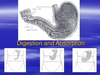
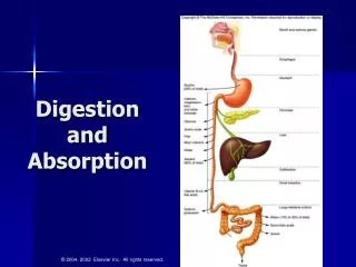
6.1 Digestion and absorption. 2. Magnification increased: intricate folded nature of the walls becomes clear. http://medcell.med.yale.edu/systems_cell_biology_old/gi/images/ small_intestine.jpg.

E N D
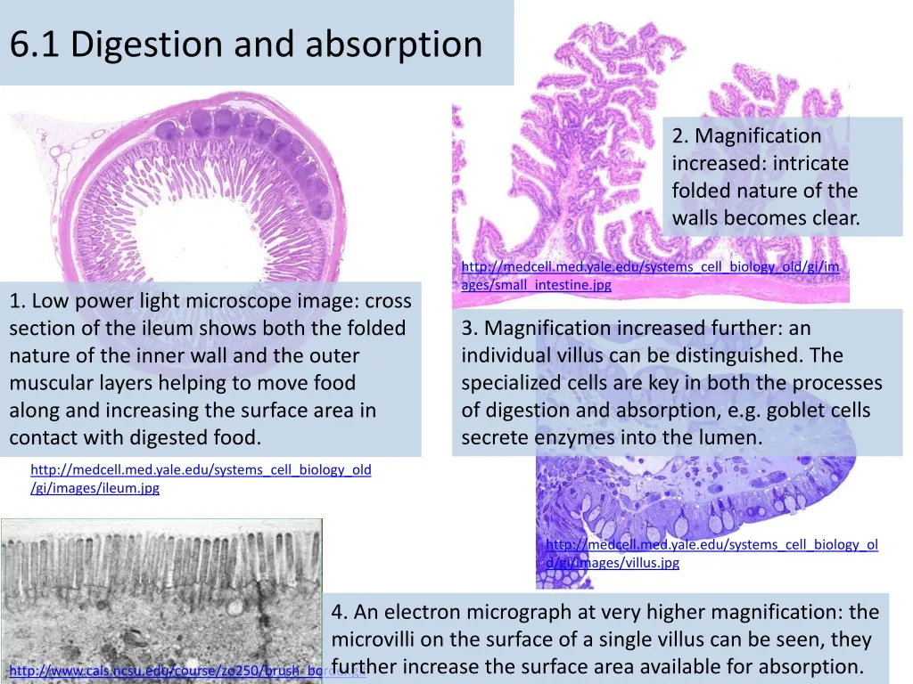
6.1 Digestion and absorption 2. Magnification increased: intricate folded nature of the walls becomes clear. http://medcell.med.yale.edu/systems_cell_biology_old/gi/images/small_intestine.jpg 1. Low power light microscope image: cross section of the ileum shows both the folded nature of the inner wall and the outer muscular layers helping to move food along and increasing the surface area in contact with digested food. 3. Magnification increased further: an individual villus can be distinguished. The specialized cells are key in both the processes of digestion and absorption, e.g. goblet cells secrete enzymes into the lumen. http://medcell.med.yale.edu/systems_cell_biology_old/gi/images/ileum.jpg http://medcell.med.yale.edu/systems_cell_biology_old/gi/images/villus.jpg 4. An electron micrograph at very higher magnification: the microvilli on the surface of a single villus can be seen, they further increase the surface area available for absorption. http://www.cals.ncsu.edu/course/zo250/brush_border.gif
6.1 • Essential idea: The structure of the wall of the intestine allows it to move, digest and absorb food.
The digestive system is fundamentally a long tube called the with two accessory organs ( ) • The role of the digestive system is: • food • Break food down into • nutrient molecules • Eliminate non-digestible remains ( )
Digestion is essential because much of the food consumed is too big to cross the gut wall and enter the blood stream • There are two types of digestion: • digestion: chopping up food and moving it along • digestion: breaking down food molecules with the use of enzymes
In order to break down large molecules of food, are needed • These enzymes are protein catalysts produced by • Enzymes make reactions happen • They greatly increase the rate at which these insoluble food substances are broken down making digestion more efficient
6.1.S1 Production of an annotated diagram of the digestive system. Use the animation and video to learn about the digestive system and how to draw it. https://youtu.be/Nm-pT7fk6gs http://highered.mheducation.com/sites/0072495855/student_view0/chapter26/animation__organs_of_digestion.html
6.1.S1 Production of an annotated diagram of the digestive system.
6.1.S1 Production of an annotated diagram of the digestive system. • Mouth: • Chewing (mechanical digestion) • Saliva moistens food to make for swallowing • Chemical digestion of produced by salivary glands
6.1.S1 Production of an annotated diagram of the digestive system. • Oesophagus: • A wave of muscle contractions ( )pushes bolus into the stomach.
6.1.S1 Production of an annotated diagram of the digestive system. • Stomach: • Muscular contractions continue . • Acid ( ) . • begins digestion of . • Thick layer of • Stomach contents are called
6.1.S1 Production of an annotated diagram of the digestive system. • Duodenum (small intestine): • from the liver and gallbladder • . • *Emulsifies fats: breaking down large globs of fat into microscopic particles increasing the surface area available for lipases (enzymes) to act on fats
6.1.S1 Production of an annotated diagram of the digestive system. • Ileum (small intestine): • Lower half of small intestine via the villi.
6.1.S1 Production of an annotated diagram of the digestive system. • Large intestine: • , leaving semi-solid feces. This is stored in the rectum.
6.1.S1 Production of an annotated diagram of the digestive system. • Egestion: • Feces (containing undigested food, dead cells and other waste) is forced out of the anus
6.1.U1 The contraction of circular and longitudinal muscle of the small intestine mixes the food with enzymes and moves it along the gut. Peristalsis moves food through the alimentary canal Contraction of . Contraction of . http://www.bbc.co.uk/schools/gcsebitesize/science/add_edexcel/common_systems/digestionrev2.shtml In the small intestine peristalsis also mixes food with enzymes and forces the products of digestion into contact with the wall of the intestine Therefore in the intestines the food is moved very slowly to allow time for digestion. n.b. The contractions are controlled unconsciously by the enteric nervous system http://www.austincc.edu/rfofi/NursingRvw/NursingPics/DigestivePics/Picture4.jpg
6.1.U3 Enzymes digest most macromolecules in food into monomers in the small intestine. • Starch, glycogen, lipids, and nucleic acids are digested into monomers • Cellulose remains undigested
Review: 2.5.U1 Enzymes have an active site to which specific substrates bind. AND2.5.U2 Enzyme catalysis involves molecular motion and the collision of substrates with the active site. Enzyme: A globular protein that increases the rate of a biochemical reaction by lowering the activation energy threshold (i.e. a biologicalcatalyst) Use the animation to find out more about enzymes and how they work. A good alternative is How Enzymes Work from McGraw and Hill http://highered.mheducation.com/sites/0072495855/student_view0/chapter2/animation__how_enzymes_work.html http://www.northland.cc.mn.us/biology/biology1111/animations/enzyme.swf
6.1.U3 Enzymes digest most macromolecules in food into monomers in the small intestine.
6.1.U3 Enzymes digest most macromolecules in food into monomers in the small intestine. Human Digestive Enzymes Remember: enzymes are specific to their substrates and each enzyme has its own optimum pH. Three main types of enzymes in human digestion: Amylases break down carbohydrates Example: Substrate: Product: Source: mouth ( ) Optimum pH: Proteases break down polypeptides Example: Substrate: Product: Source: Optimum pH: Lipases break down fats and lipids Example: Substrate: Product: Source: Optimum pH: diagram from: http://www.teachervision.fen.com/digestive-system/printable/57730.html
6.1.U2 The pancreas secretes enzymes into the lumen of the small intestine. The synthesizesthe three main types of digestive enzyme: • to digest carbohydrates, e.g. starch • to digest lipids, e.g. triglycerides • to digest polypeptides Pancreatic juice containing the enzymes is released into the upper region of the small intestine (duodenum) via the The small intestine is where the final stages of digestion occur. https://commons.wikimedia.org/wiki/File:Diagram_showing_the_position_of_the_pancreas_CRUK_356.svg
6.1.S2 Identification of tissue layers in transverse sections of the small intestine viewed with a microscope or in a micrograph. The small intestine contains four distinct tissue layers from the lumen: 1. Mucosa– 2. Submucosa– connective tissue ( ) 3. Muscular layer – and muscle perform peristalsis 4. Serosa– Epithelial cells – (see 6.1.U4) Muscular layer circular longitudinal *You should be able to label: longitudinal & circular muscles, mucosa and epithelium http://www.dartmouth.edu/~anatomy/Histo/lab_5/GI/DMS132/popup.html
6.1.U4 Villi increase the surface area of epithelium over which absorption is carried out. Adaptations to Absorption Getting digested food molecules into the blood from the lumen of the ileum. Many protrude into the lumen, greatly increasing the . Single-cell layer of epithelial cells . on the surface of each cell . Lacteals (lymph vessels) Allow for . Capillaries close to epithelium Short path for diffusion, rich supply of blood. Rich blood supply Maintains concentration gradients between lumen and blood. Images from: http://en.wikipedia.org/wiki/Intestinal_villi
6.1.U5 Villi absorb monomers formed by digestion as well as mineral ions and vitamins. https://youtu.be/P1sDOJM65Bc Along with vitamins and minerals all products of digestion (monosaccharides, amino acids, fatty acids & glycerol) are absorbed by the villi
6.1.U6 Different methods of membrane transport are required to absorb different nutrients. How is membrane transport involved in absorption of nutrients from the small intestine?
6.1.A1 Processes occurring in the small intestine that result in the digestion of starch and transport of the products of digestion to the liver. Starch consists of (by ) and (by ) Amylase breaks Maltase Dextrinase breaks the http://etravelweek.com/hmattachments/1_200907180843167cXQr.gif
6.1.A1 Processes occurring in the small intestine that result in the digestion of starch and transport of the products of digestion to the liver. The digested glucose is absorbed and then transported to various body tissues Glucose is (of the villus). Glucose moves by . Glucose then diffuses a short distance into the . Blood in the capillaries moves to to. The liver absorbs excess glucose which it converts to for storage. http://www.rpi.edu/dept/chem-eng/Biotech-Environ/Membranes/bauerp/co.gif
6.1.A2 Use of dialysis tubing to model absorption of digested food in the intestine. Dialysis (visking) tubing can be used to model absorption The tubing is semi-permeable and contains pores typically ranging 1 – 10 nm in diameter Predict what will happen to the glucose and starch after 15 minutes. Initially contains a mixture of starch and glucose Test the solutions inside and outside the dialysis tubing for starch and glucose before and after at least 15 minutes have elapsed(see the Practical Biology link for details). http://www.nuffieldfoundation.org/practical-biology/evaluating-visking-tubing-model-gut
6.1.A2 Use of dialysis tubing to model absorption of digested food in the intestine. Dialysis (visking) tubing can be used to model absorption The tubing is semi-permeable and contains pores typically ranging 1 – 10 nm in diameter Predict what will happen to the glucose and starch after 15 minutes. • The model is the most basic element of the scientific method. It is any simplification, substitute or stand-in for what you are actually studying or trying to predict. Evaluate the usefulness of dialysis tubing as a model for absorption by considering: • How is the function of dialysis tubing similar to the small intestine? • What features of a real gut are missing from this model? Initially contains a mixture of starch and glucose Test the solutions inside and outside the dialysis tubing for starch and glucose before and after at least 15 minutes have elapsed(see the Practical Biology link for details). http://www.nuffieldfoundation.org/practical-biology/evaluating-visking-tubing-model-gut