Download
1 / 19
280 likes | 730 Views
FOREARM. By Prof. Saeed Abuel Makarem Dr. Sanaa Al-Sharawy. By the end of the lecture the student should be able to: Enumerate the different muscles of the front (flexors) and back (extensors) of the forearm. Describe in brief the attachment of these group of muscles:

E N D
FOREARM By Prof. Saeed Abuel Makarem Dr. Sanaa Al-Sharawy
By the end of the lecture the student should be able to: • Enumerate the different muscles of the front (flexors) and back (extensors) of the forearm. • Describe in brief the attachment of these group of muscles: • Superficial and deep flexors; • Superficial and deep extensors. • Describe the action and nerve supply of each of these muscle. OBJECTIVES
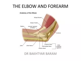
The forearm extends from elbow to wrist. • It posses two bones radius laterally & Ulna medially. • The two bones are connected together by the interosseous membrane. • This membrane allows movement of Pronation and Supination while the two bones are connected together. • Also it gives origin for the deep muscles.
The forearm is enclosed in a sheath of deep fascia, which is attached to the post. subcutaneous border of the ulna . This fascial sheath, together with the interosseous membrane, radius & ulna and a fibrous intermuscular septum divides the forearm into several compartments. Each compartments have its own muscles, nerves, and blood supply. Fascial Compartments of the Forearm
Anterior compartment -FLEXOR GROUP • These muscles: are (8) • They act on the wrist & elbow joints and the fingers. • They form fleshy masses in the proximal forearm and become tendinous in the distal part of the forearm. • They are arranged in three groups: I-Superficial: 4 • Pronator teres • Flexor carpi radialis • Palmaris longus • Flexor carpi ulnaris II-Intermediate: 1 • Flexor digitorum superficialis III- Deep: 3 • Flexor digitorum profundus. U • Flexor pollicis longus. R • Pronator quadratus. R & U
Superficial Flexors • They arise - more or less- from the common flexor origin (front of medial epicondyle). • All are supplied by the median nerve exceptone,flexor carpi ulnaris, FCU (ulnar n.). • All cross the wrist joint except one, pronator teres, (PT).
Flexor Carpi Radialis • Insertion: Base of 2nd metacarpal bone • Action: Flexion & abduction of the wrist. • Palmaris Longus • Insertion: into the flexor retinaculum & palmer aponeurosis. • Action: Flexes hand & tightens the palmer aponeurosis • Pronator teres Insertion: middle of lat. surface of radius • Action: pronation & flexion of forearm . May Be Absent
Flexor Digitorum Superficialis • Origin: • Common flexor origin, • Coronoid process of ulna; • Anterior oblique line of radius • Insertion: • base of middle phalanges of the medial 4 fingers. • Action: • Flexes middle and proximal phalanges of medial 4 fingers • Flexes the hand (wrist). • Flexor Carpi Ulnaris • Insertion: • Pisiform, • hook of hamate • 5th metacarpal bone • Action: • Flexion and adduction of the hand (wrist)
Deep Flexors • One above radius: Flexor pollicis longus • One above ulna: Flexor Digitorum profundus • One above the two bones: Pronator Quadratus.
Pronator Quadratus • Insertion: distal one fourth of ant. surface of radius • Action: pronates the forearm (primover), • Hold the two bones together • Flexor Pollicis Longus • Insertion: Base of distal phalanx of thumb • Action: flexes (all joints of the thumb), interphalangeal, metacarpophalangeal & carpometacarpal joints. • Flexor Digitorum Profundus • Insertion: bases of the distal phalanges of the medial four digits • Action: Flexes distal phalanges of medial four digits
Supination and pronation It occurs in the superior and inferior radioulnar joints; (pivot synovial joint) Muscles produce supination Biceps brachii. Supinator. Muscles produce pronation Pronator teres. pronator quadratus. NB. Brachioradialis put the forearm in midprone-supine position, (initiates pronation and supination).
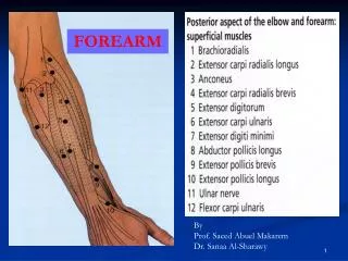
Posterior Compartment: 3 groups • Deep group 5 (3 to thumb+ 1 to index + supinator). • Supinator. • Abductor pollicis longus. • Extensor pollicis brevis. • Extensor pollicis longus. • Extensor indices. Lateral group 2 Brachioradialis. Extensor carpi radialis longus. (These two muscles arises from the lateral supracondylar ridge). Superficial group 5 Extensor carpi radialis brevis. Extensor digitorum . Extensor digiti minimi. Extensor carpi ulnaris. Anconeus . Origin: Common Extensor Origin (front of the lateral epicondyle).
Posterior compartment • I- Superficial group: • 7 muscles ( from lateral to medial) • Brachioradialis, (BR). • Extensor carpi radialis longus, (ECRL). • Extensor carpi radialis brevis, (ECRB). • Extensor digitorum, (ED). • Extensor digiti minimi, (EDM). • Extensor carpi ulnaris, (ECU). • Anconeus. (An).
Superficial extensor • All arises from the commonextensor origin, (front of lateral epicondyle of the humerus), EXCEPT, 2 (BR & EXRL). • All cross the wrist EXCEPT, one, (brachioradialis. • All supplied by deep branch of radial nerve, EXCEPTABE • A, Anconeus • B, Brachioradialis • E, Extensor carpi radialis longus • These 3 muscles are supplied by the radial nerve itself
Extensor Carpi radialis longus • Origin: • Lateral supracondylar ridge of humerus • Insertion: • Posterior surface of base of second metacarpal bone • Action: • Extends and abducts hand at wrist joint • Brachioradialis • Origin: • Lateral supracondylar ridge of humerus • Insertion: • Base of styloid process of radius • Action: • Flexes forearm; (elbow). • Rotates forearm to the midprone position
INSERTION Extensor carpi radialis brevis: base of 3rd metacarpal bone. Extensor digitorum: Extensor expansion of the medial 4 fingers. Extensor digiti minimi: Extensor expansion of the little finger. Extensor carpi ulnaris: Base of the 5th metacarpal bone.
II- Deep group: 5 muscles 1- Abductor pollicis longus, (APL). 2- Extensor pollicis brevis, (EPB). 3- Extensor pollicis longus, (EPL). 4- Extensor indicis (EI). 5- Supinator. All back muscles of forearm are supplied by posterior interosseous nerve except , ABE by Radial nerve.
Dorsal Extensor Expansion It is formed by the union of the tendons of: Extensor digitorum, Extensor indicis, extensor digiti minimi, palmar interossei, dorsal interossei and lumbricals muscles. All these tendons unite to form one tendon which divides into 3 slips, a median one attached to middle phalanges and 2 lateral attached to the terminal phalanges.