Download
1 / 31
420 likes | 980 Views
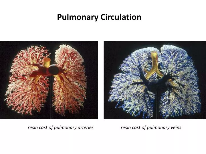
Pulmonary Circulation. resin cast of pulmonary arteries resin cast of pulmonary veins. pulmonary circulation . Pulmonary Circulation The pulmonary circulation is unique in a number of ways.

E N D
Pulmonary Circulation resin cast of pulmonary arteries resin cast of pulmonary veins
Pulmonary Circulation The pulmonary circulation is unique in a number of ways. 1. The pulmonary circulation has to carry the entire cardiac output – unlike most other organ systems which only carry a fraction of total output. 2. Because the lung has to deal with the entire cardiac output – it has a high capacity, but because the lung is such a fragile network (blood gas barrier is a fraction of a micron) it has to avoid high pressure. 2. Dealing with the entire cardiac output, the lung is uniquely positioned to condition blood – many blood components are metabolized by the pulmonary vascular endothelium as they go through the lung. It also places the lung in the unique position of being able to trap thrombi and emboli. Metabolized blood components e.g.- prostaglandin E1, E2 and F2a, leukotrienes, serotonin, acetylcholine norepiphrine , Bradykininare all metabolized. angiotensinI - 70% conversion to AngiotensinII.
Comparison of Vascular Pressures in the Systemic & Pulmonary Circulations • 10 fold difference in mean arterial pressure • structural basis: less smooth muscle in pulmonary vessels - greater distensibility + greater compressibility • major drop in pressure in the pulmonary circulation is through capillaries • major drop in pressure in the systemic circulation is through the arterioles Low Pressure High Capacity System It is imperative that pulmonary blood pressure is maintained below 25 mm Hg, as an increase above this level represents sufficient hydrostatic pressure to ‘squeeze’ water out of the vascular space into the extracellular space or even the alveoli themselves.
Right Heart Catheterization: Measuring R-side Pressures Pulmonary Vascular Resistance PVR = ΔP/ΔQ = PPA – PLA / C.O. = 15 – 5 / 5 = 2 mmHg/L/min A Swan-Ganz catheter introduced through a peripheral vein(femoral/ brachial/ jugular), advanced toward the chest by normal flood flow, allows for RA, RV & pulmonary artery “wedge” [estimates LA] pressures.
Capillary Network vs. Capillary Bed Pulmonary capillary beds are distinct from systemic capillaries. Systemic capillaries have a small diameter and represent a high resistance to blood flow. The pulmonary capillary beds are essentially a ‘sheet’ of blood that flows across the alveolar surface. This arrangement has less resistance to flow. Recruitment and distension of vessels allows flow to increase in the pulmonary vessels whilst resistance DECREASES
Recruitment – closed vessels or vessels with no flow now conduct blood, i.e. more area for flow is opened up.
Distention – the vessel walls dilate and the cross sectional area increases – reducing resistance. An increase in cardiac output results in both recruitment and distension of pulmonary vessels and therefore the pulmonary vascular resistance falls. vascular resistance (R) is equal to the pressure gradient (ΔP) divided by blood flow Significance: pulmonary circulation can accommodate a large increase in cardiac output with only a small increase in pulmonary arterial pressure
Passive Influences on PVR Distention & Recruitment Increase in perfusion pressure [pulmonary artery pressure] results in distension & recruitment ⇒ decreasing PVR. How can vessels be open but have no flow? Consider very low pressure systems, e.g. garden hose with multiple small holes. At low enough pressure, only a few holes drizzle water: sufficient difference in resistance that flow is diverted to the path with least resistance
The fall in pulmonary vascular resistance with increased cardiac output has two beneficial effects:- -> It opposes the tendency of blood velocity to speed up with increased flow rate, maintaining adequate time for pulmonary capillary blood to take up oxygen and dispose of carbon dioxide. ->It also results in an increase in capillary surface area, which enhances the diffusion of oxygen into and carbon dioxide out of the pulmonary capillary blood. Capillary recruitment and distention also have a protective function. High capillary pressure is a major threat to the lungs and can cause pulmonary edema, an abnormal accumulation of fluid, which can flood the alveoli and impair gas exchange. When cardiac output increases from a resting level of 5 L/min to 25 L/min with vigorous exercise, the decrease in pulmonary vascular resistance not only minimizes the load on the right heart but also keeps the capillary pressure low and prevents excess fluid from leaking out of the pulmonary capillaries
The left slide shows that increasing cardiac output (as measured by the pressure in the pulmonary artery immediately after blood exits the heart) results in a drop of pulmonary vascular resistance. This is important since recruitment and distention of pulmonary vessels allows an increase in flow due to a fall in resistance. Furthermore, the lowered PVR means that only small changes in pressure are required to achieve large changes in flow. The right slide shows that a mere 5 mm Hg increase in pulmonary pressure produces a quadrupling of flow through the lung.
Passive Influences on PVR Difference in Surrounding Pressure • Alveolar vessels [pulmonary capillaries] – alveolar pressure • Extra-alveolar vessels [pulmonary arteries & veins]- intrapleural pressure Lung inflation: • collapses alveolar vessels via stretch of alveolar wall • expands extra-alveolar vessels via radial traction
Lung Volume Affects Pulmonary Vascular Resistance Residual vol= Alveolar low resistance Extra-Alveolar – high resistance FRC = both vessels resistance relatively low Total lung Cap. = Alveolar resistance high Extra-alveolar resistance low Therefore – total pulmonary vascular resistance, i.e. the sum of alveolar vessel + extra-alveolar vessel resistance (lower graph) is lowest when the lung is at FRC.
Active influences on Pulmonary Vascular Resistance 1. Hypoxia – increases PVR – in order to maintain blood flow through oxygenated areas of lung hypoxia causes vasoconstriction of pulmonary vessels in poorly oxygenated areas of lung and a local increase in PVR. This can be highly localized to specific areas of lung (i.e. poorly ventilated areas) or in the case of general hypoxia (e.g. at altitude) throughout the pulmonary vasculature. Mechanism for this hypoxic increase in PVR is still unknown. An increase in PVR will reduce blood flow – 2. Serotonin, histamine and norepinephrin(Humoral) increase PVR – smooth muscle contraction. 3. Isoproteranol, nitric oxide and acetylcholine(Humoral) decrease PVR – smooth muscle relaxation. There is sparse sympathetic & parasympathetic innervation of the pulmonary vasculature and the effect of stimulation of these nerves is controversial
Summary • Consider the factors that affect pulmonary vascular resistance (PVR). How do these differ from factors that affect systemic vascular resistance (TPR) • Contrast the effect of low oxygen on vessel diameter in the pulmonary versus systemic circulation. How is this difference useful? When is it not beneficial?
16 Blood Flow Distribution in the Lungs The effects of gravity on blood flow are dramatic and result in an uneven distribution of blood in the lungs. In an upright person, the gravitational pull on the blood is downward. Because the vessels are highly compliant, gravity causes the blood volume and flow to be greater at the bottom of the lung (the base) than at the top (the apex). Against Gravity With Gravity Blood Flow
The uneven distribution of blood flow can be explained by the hydrostatic pressure differences within the blood vessels. • If we consider the pulmonary arterial system as a continuous column of blood, the difference in pressure between the top and bottom of a lung 30 cm high will be about 30 cm water, or 23 mm Hg. • This is a large pressure difference for such a low-pressure system as the pulmonary circulation • When considering the pressures in the pulmonary vasculature an artificial (theoretical) categorization can be used. In practice regional lung blood flow is a highly variable parameter – however, this zone categorization is a useful tool to think about pulmonary vasculature and pressures.
The “Zones” of the Lung: Explanation of the uneven distribution of blood flow in the lung, based on the pressures affecting the capillaries. zone 1 - a region at the top of the lung where pulmonary arterial pressure falls below alveolar pressure. If this occurs, the capillaries are squashed flat, and no flow is possible. Zone 1 does not occur under normal conditions, because the pulmonary arterial pressure is just sufficient to raise blood to the top of the lung, but may be present if the arterial pressure is reduced (following severe hemorrhage, for example) or if alveolar pressure is raised (during positive pressure ventilation). This ventilated but unperfused lung is useless for gas exchange and is called alveolar dead space. pressure gradient in zone 1 is represented as PA > Pa > Pv
zone 2 condition occurs in the middle of the lungs, where pulmonary arterial pressure, caused by the increased hydrostatic effect, is greater than alveolar pressure . • Venous pressure is less than alveolar pressure. • As a result, blood flow in a zone 2 condition is determined not by the arterial-venous pressure difference, but by the difference between arterial pressure and alveolar pressure. • The pressure gradient in zone 2 is represented as Pa > PA > Pv. • The functional importance of this is that venous pressure in zone 2 has no effect on flow (i.e., lowering venous pressure will not increase capillary blood flow in this zone).
zone3- venous pressure now exceeds alveolar pressure, and flow is determined in the usual way by the arterial-venous pressure difference. • The increase in blood flow down this region of the lung is apparently caused chiefly by distension of the capillaries. The pressure within them (lying between arterial and venous) increases down the zone while the pressure outside (alveolar) remains constant. • Thus, their transmural pressure rises and, indeed, measurements show that their mean width increases. Recruitment of previously closed vessels may also play some part in the increase in blood flow down this zone.
Zone Summary PA > PPa > PPv Zone I no flow PPa > PA > PPv Zone II restricted flow flow driven by PPa – PA difference PPa > PPv > PA Zone III highest flow flow driven by PPa – PPv difference Note that as we move down the Zones we simply move PA one step to the right (and of course PPa is always going to be higher than PPv).
Gravity Causes a Mismatch of Regional Ventilation and Blood Flow in the Lungs- Even though total ventilation and total blood flow (i.e., cardiac output) may be normal, there are regions in the lung where ventilation and blood flow are not matched, so that a certain fraction of the cardiac output is not fully oxygenated. The matching of airflow and blood flow is best examined by considering the ventilation,perfusion ratio, which compares alveolar ventilation with blood flow in lung regions. Because resting healthy people have an alveolar ventilation ([V with dot above]A) of 4 L/min and a cardiac output ([Q with dot above]) of 5 L/min, the ideal alveolar ventilation,perfusion ratio ([V with dot above]A/[Q with dot above]) should be 0.8 (there are no units, as this is a ratio).
We have already seen that gravity can cause regional differences in blood flow and alveolar ventilation. In an upright person, the base of the lungs is better ventilated and better perfused than the apex. Regional alveolar ventilation and blood flow are illustrated in Figure . Three points are apparent from this figure: • Ventilation and blood flow are both gravity-dependent, airflow and blood flow increase down the lung. • Blood flow shows about a five-fold difference between the top and bottom of the lung, whereas ventilation shows about a two-fold difference. This causes gravity-dependent regional variations in the ventilation,perfusion (VA/Q)ratio that range from 0.6 at the base to 3 or higher at the apex. • Blood flow is proportionately greater than ventilation at the base, and ventilation is proportionately greater than blood flow at the apex.
The functional importance of lung ventilation,perfusionratios is in terms of gas exchange. • At the apical region, where the V A/Q ratio is higher than 0.8, there is overventilation relative to blood flow. • At the base, where the ratio is lower than .08, the opposite occurs (i.e., overperfusion relative to ventilation). • In the latter case, a fraction of the blood passes through the pulmonary capillaries at the base of the lungs without becoming fully oxygenated. Regional differences in V,Qratiostend to localize some diseases to the top or bottom parts of the lungs. For example, tuberculosis tends to be localized in the apex because of a more favorable environment (i.e., higher oxygen levels for Mycobacterium tuberculosis). • Because overventilation relative to blood flow occurs in the apex, the PaO2 is high and the PaCO2 is low at the apex of the lungs
Shunts and Venous Admixture • Matching of the lungs' airflow and blood flow is not perfect. • On one side of the alveolar-capillary membrane there is Wasted air(i.e., physiological dead space), and on the other side there is wasted blood. Wasted blood refers to any fraction of the venous blood that does not get fully oxygenated. • The mixing of unoxygenated blood with oxygenated blood is known as venous admixture. • Two causes for venous admixture: • -a shunt • -and a low V A/Q ratio. 1. An anatomic shunt -right-to-left shunt -bronchial circulation 2. low regional ventilation,perfusion ratio -In a healthy person, a low V A/Q ratio occurs at the base of the lung - can also occur with a partially obstructed airway
In summary, venous admixture results from anatomic shunt and a low regional V A/Q ratio. • In healthy people, approximately 50% of the venous admixture comes from an anatomic shunt (e.g., bronchial circulation) and 50% from a low [V with dot above]A/[Q with dot above] ratio at the base of the lungs as a result of gravity. • Physiological shunt (i.e., total venous admixture) represents about 1% to 2% of cardiac output in healthy people. • This amount can increase up to 15% of cardiac output with some bronchial diseases, and, in certain congenital disorders, a right-to-left anatomic shunt can account for up to 50% of cardiac output. • A good way to remember the importance of a shunt is that it always leads to venous admixture and reduces the amount of oxygen carried in the systemic blood.
Pulmonary Edema Pulmonary edema will develop when the net balance of forces acting on water transport favors the movement of fluid from the vascular space into the extracellular space or even the alveolar space. The main factors influencing water transport in this space are 1. Hydrostatic pressure, i.e. blood pressure in the capillaries – usually 8-10 mm Hg 2. Oncotic pressure of the blood – i.e. the osmotic pressure of plasma proteins in blood – about 25 mm Hg 3. Alveolar pressure – strong +ve pressure push water into extracellular space from alveolar space (not usually an issue) 4. Surface tension – surface tension pulls on the alveolus and decreases the hydrostatic pressure in the extracellular space – causing water to be sucked into that space. Can also act to pull water into the alveolar space. 5. Lymphatic drainage – there is a net movement of fluid from the vascular to the extracellular space of 20 ml hour. This is cleared by the lymphatic system.