Download
1 / 1
10 likes | 91 Views
A. B. AMKL11 stained CD7/33. AMKL11 sorted blasts. Supplementary Figure 2: Cell sorting Patient samples (example in A and B)

E N D
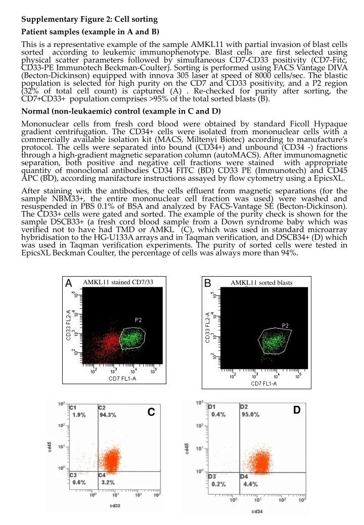
A B AMKL11 stained CD7/33 AMKL11 sorted blasts Supplementary Figure 2: Cell sorting Patient samples (example in A and B) This is a representative example of the sample AMKL11 with partial invasion of blast cells sorted according to leukemic immunophenotype. Blast cells are first selected using physical scatter parameters followed by simultaneous CD7-CD33 positivity (CD7-Fitc, CD33-PE Immunotech Beckman-Coulter). Sorting is performed using FACS Vantage DIVA (Becton-Dickinson) equipped with innova 305 laser at speed of 8000 cells/sec. The blastic population is selected for high purity on the CD7 and CD33 positivity, and a P2 region (32% of total cell count) is captured (A) . Re-checked for purity after sorting, the CD7+CD33+ population comprises >95% of the total sorted blasts (B). Normal (non-leukaemic) control (example in C and D) Mononuclear cells from fresh cord blood were obtained by standard Ficoll Hypaque gradient centrifugation. The CD34+ cells were isolated from mononuclear cells with a commercially available isolation kit (MACS, Miltenyi Biotec) according to manufacture’s protocol. The cells were separated into bound (CD34+) and unbound (CD34 -) fractions through a high-gradient magnetic separation column (autoMACS). After immunomagnetic separation, both positive and negative cell fractions were stained with appropriate quantity of monoclonal antibodies CD34 FITC (BD) CD33 PE (Immunotech) and CD45 APC (BD), according manifacture instructions assayed by flow cytometry using a EpicsXL. After staining with the antibodies, the cells effluent from magnetic separations (for the sample NBM33+, the entire mononuclear cell fraction was used) were washed and resuspended in PBS 0.1% of BSA and analyzed by FACS-Vantage SE (Becton-Dickinson). The CD33+ cells were gated and sorted. The example of the purity check is shown for the sample DSCB33+ (a fresh cord blood sample from a Down syndrome baby which was verified not to have had TMD or AMKL (C), which was used in standard microarray hybridisation to the HG-U133A arrays and in Taqman verification, and DSCB34+ (D) which was used in Taqman verification experiments. The purity of sorted cells were tested in EpicsXL Beckman Coulter, the percentage of cells was always more than 94%. D C