Download
1 / 93
950 likes | 1.31k Views
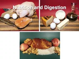
Digestion and Nutrition. Introduction. Digestion refers to the mechanical and chemical breakdown of foods so that nutrients can be absorbed by cells. The digestive system carries out the process of digestion.

E N D
Introduction • Digestion refers to the mechanical and chemical breakdown of foods so that nutrients can be absorbed by cells. • The digestive system carries out the process of digestion. • The digestive system consists of the alimentary canal, leading from mouth to anus, and several accessory organs whose secretions aid the processes of digestion.
I. General Characteristics of the Alimentary Canal • The alimentary canal is a muscular tube about 9 meters long that passes through the body's ventral cavity.
Structure of the Wall • Structure of the Wall • The wall of the alimentary canal consists of the same four layers throughout its length, with only slight variations according to the functions of specific sections of the canal. • The inner layer is the mucosa, which is lined with epithelium attached to connective tissue; it protects tissues of the canal and carries on secretion and absorption.
Structure of the Wall cont. • The next layer is the submucosa, which is made up of loose connective tissue housing blood and lymph vessels and nerves; it nourishes the surrounding layers of the canal. • The muscular layer consists of inner circular fibers and outer longitudinal fibers that propel food through the canal. • The outer layer, or serosa, is composed of visceral peritoneum that protects underlying tissues and secretes serous fluid to keep the canal from sticking to other tissues in the abdominal cavity.
Movements of the Tube • Movements of the Tube • The motor functions of the alimentary canal are of two types— mixing movements and propelling movements. • Mixing movements occur when smooth muscles contract rhythmically in small sections of the tube. • Propelling movements include a wavelike motion called peristalsis, which is caused by contraction behind a mass of food as relaxation allows the mass to enter the next segment of the tube.
II. Mouth • The mouth is the first portion of the alimentary canal; it functions to receive food and begins mechanical digestion by mastication.
Cheeks and Lips • Cheeks and Lips • Cheeks form the lateral walls of the mouth. • The lips are highly mobile structures that surround the mouth opening. • The lips contain sensory receptors that help to judge the temperature and texture of food.
Tongue • Tongue • The tongue is a thick, muscular organ covered by mucous membrane with taste buds within papillae; it is attached to the floor of the mouth by the frenulum. • The papillae also provide friction for moving food around in the mouth. • Lingual tonsils are lymphatic tissues located at the root of the tongue.
Palate • Palate • The palate forms the roof of the oral cavity and has an anterior hard palate and posterior soft palate. • The soft palate and uvula function to close off the nasal cavity during swallowing. • Associated with the palate in the back of the mouth are palatine tonsils, which, because they are lymphatic tissue, help to protect the body against infection. • Another lymphatic tissue mass, pharyngeal tonsils (adenoids), are located on the posterior wall of the pharynx, above the border of the soft palate.
Teeth • Teeth • Two sets of teeth develop in sockets within the alveolar processes of the maxillary and mandibular bones. • The 20 primary teeth are shed in the order they appeared and are replaced by 32 secondary teeth. • Through the actions of chewing, teeth break food into smaller pieces, beginning mechanical digestion.
Teeth cont. • Different teeth are adapted to handle food in different ways, and include incisors, cuspids, bicuspids, and molars. • Each tooth consists of a crown and a root, and is made of enamel, dentin, pulp, cementum, nerves, and blood vessels. • A tooth is held tight in its socket by a periodontal ligament.
III. Salivary Glands • The salivary glands secrete saliva, which moistens and dissolves food particles, binds them together, allows tasting, helps to cleanse the mouth and teeth, and begins carbohydrate digestion.
Salivary Secretions • Salivary Secretions • Salivary glands contain serous cells that produce a watery fluid with amylase, and mucous cells that produce lubricating and binding mucus. • Salivary glands receive parasympathetic stimulation that triggers the production of a large volume of saliva at the sight or smell of food.
Major Salivary Glands • Major Salivary Glands • The parotid glands, lying in front of the ear, are the largest of the major salivary glands; they secrete a clear, watery fluid rich in amylase. • The submandibular glands, located on the floor of the mouth, secrete a more viscous fluid. • The sublingual glands, inferior to the tongue, are the smallest of the major salivary glands and secrete a saliva that is thick and stringy.
IV. Pharynx & Esophagus • The pharynx is a cavity lying behind the mouth, and the esophagus is a muscular tube leading to the stomach.
Structure of the Pharynx • Structure of the Pharynx • The pharynx connects the nasal and oral cavities with the larynx and esophagus and is divided into a nasopharynx (top portion), oropharynx (middle portion), and largyngopharynx (bottom portion).
Swallowing Mechanism • Swallowing Mechanism • Swallowing reflexes can be divided into three stages. • Food is mixed with saliva and voluntarily forced into the pharynx with the tongue. • Sensory receptors in the pharynx sense food, which triggers swallowing reflexes. • In the third stage of swallowing, peristalsis transports the food in the esophagus to the stomach.
Esophagus • Esophagus • The esophagus is a straight, collapsible passageway leading to the stomach. • Mucous glands are scattered throughout the submucosa of the esophagus and produce mucus to moisten and lubricate the inner lining of the tube. • The lower esophageal sphincter helps to prevent regurgitation of the stomach contents into the esophagus.
V. Stomach • The stomach is a J-shaped muscular organ that receives and mixes food with digestive juices, and propels food to the small intestine.
Parts of the Stomach • Parts of the Stomach • The stomach is divided into cardiac, fundic, body, and pyloric regions and a pyloric canal. • The pyloric sphincter controls release of food from the stomach into the small intestine.
Gastric Secretions • Gastric Secretions • Gastric glands within the mucosa of the stomach open as gastric pits. • Gastric glands generally contain three types of secretory cells. • Mucous cells produce mucus that protects the stomach lining.
Gastric Secretions cont. • Chief cells secrete pepsin (to digest protein) as inactive pepsinogen, which is activated when it comes in contact with hydrochloric acid. • Parietal cells secrete hydrochloric acid. • Other components of gastric juice include intrinsic factor, required for vitamin B12 absorption from the small intestine.
Regulation of Gastric Secretions • Regulation of Gastric Secretions • Gastric secretions are enhanced by parasympathetic impulses and the hormone gastrin, which is released from gastric glands. • As more food enters the small intestine, secretion of gastric juice from the stomach wall is reflexly inhibited. • Presence of fats and proteins in the upper small intestine causes the release of cholecystokinin from the intestinal wall, which also decreases gastric mobility.
Gastric Absorption • Gastric Absorption • The stomach absorbs only small quantities of water and certain salts, alcohol, and some lipid-soluble drugs.
Mixing and Emptying Actions • Mixing and Emptying Actions • Following a meal, mixing actions of the stomach turn the food into chyme and pass it toward the pyloric region using peristaltic waves. • The rate at which the stomach empties depends on the fluidity of the chyme and the type of food. • As chyme fills the duodenum, stretching of its wall triggers the enterogastric reflex, which inhibits peristalsis and slows the rate at which chyme enters the small intestine.
VI. Pancreas • The pancreas has an exocrine function of producing pancreatic juice that aids digestion.
Structure of the Pancreas • Structure of the Pancreas • The pancreas is closely associated with the small intestine. • The cells that produce pancreatic juice, called pancreatic acinar cells, make up the bulk of the pancreas. • Pancreatic acinar cells cluster around tiny tubes that merge to form larger ones, and then give rise to the pancreatic duct. • The pancreatic and bile ducts join and empty into the small intestine, which is surrounded by the hepatopancreatic sphincter.
Pancreatic Juice • Pancreatic Juice • Pancreatic juice contains enzymes that digest carbohydrates, fats, proteins, and nucleic acids. • Pancreatic enzymes include pancreatic amylase, pancreatic lipase, trypsin, chymotrypsin, carboxypeptidase, and two nucleases. • Protein-digesting enzymes are released in an inactive form and are activated upon reaching the small intestine.
Regulation of Pancreatic Secretion • Regulation of Pancreatic Secretion • The nervous and endocrine systems regulate release of pancreatic juice. • Secretin from the duodenum stimulates the release of pancreatic juice with a high bicarbonate ion concentration but few digestive enzymes. • Cholecystokinin from the wall of the small intestine stimulates the release of pancreatic juice with abundant digestive enzymes.
VII. Liver • The reddish-brown liver, located in the upper right quadrant of the abdominal cavity, is the body’s largest internal organ.
Liver Structure • Liver Structure • The liver is divided into right and left lobes, and is enclosed by a fibrous capsule. • Each lobe is separated into hepatic lobules consisting of hepatic cells radiating from a central vein.
Liver Structure cont. • Hepatic sinusoids separate groups of hepatic cells. • Blood from the hepatic portal vein carries blood rich in nutrients to the liver. • Kupffer cells carry on phagocytosis in the liver. • Secretions from hepatic cells are collected in bile canals that converge to become hepatic ducts and finally form the common hepatic duct.
Liver Functions • Liver Functions • The liver carries on many diverse functions for the body. • The liver is responsible for many metabolic activities, such as the metabolism of carbohydrates, lipids, and proteins. • The liver also stores glycogen, vitamins A, D, and B12, iron, and blood.
Liver Functions cont. • The liver filters the blood, removing damaged red blood cells and foreign substances, and removes toxins. • The liver's role in digestion is to secrete bile.
Composition of Bile • Composition of Bile • Bile is a yellowish-green liquid that hepatic cells secrete; it includes water, bile salts, bile pigments, cholesterol, and electrolytes. • Bile pigments are breakdown products from red blood cells. • Only the bile salts have a digestive function.
Gallbladder • Gallbladder • The gallbladder is a pear-shaped sac lying on the interior surface of the liver. • It is connected to the cystic duct, which joins the hepatic duct; these two ducts merge to form the common bile duct leading to the duodenum. • A sphincter muscle controls the release of bile from the common bile duct.
Regulation of Bile Release • Regulation of Bile Release • Bile does not normally enter the duodenum until cholecystokinin stimulates the gallbladder to contract. • The hepatopancreatic sphincter remains contracted unless a peristaltic wave approaches it, at which time it relaxes and a squirt of bile enters the duodenum.
Functions of Bile Salts • Functions of Bile Salts • Bile salts emulsify fats into smaller droplets and aid in the absorption of fatty acids, cholesterol, and certain vitamins.