Download
1 / 24
290 likes | 752 Views
Regulation of MHC II Gene Transcription. Charlotte S. Kaetzel PhD Dept. of Microbiology, Immunology & Molecular Genetics February 28, 2008. MHC II Function. Expressed by antigen-presenting cells (APCs) Heterodimer of a and b chains Binds small peptides “Presents” peptides to T cells.

E N D
Regulation of MHC II Gene Transcription Charlotte S. Kaetzel PhD Dept. of Microbiology, Immunology & Molecular Genetics February 28, 2008
MHC II Function • Expressed by antigen-presenting cells (APCs) • Heterodimer of a and b chains • Binds small peptides • “Presents” peptides to T cells MHC II TCR
Constitutive “Professional” APCs B cells Dendritic cells Inducible by IFN-g or LPS Many cell types Macrophages Epithelial cells Fibroblasts Others Expression of MHC II Molecules Down-regulated in B cell → plasma cell differentiation
MHC II a chain genes MHC II pseudogenes MHC II b chain genes unrelated genes Organization of MHC II genes Short arm of human chromosome 6
MHC II Gene Expression • Heterodimers of a and b subunits • Coordinate expression of multiple loci: DR, DQ, DO, DM, DP • Co-dominant expression of maternal and paternal alleles
The MHC II Promoter Nekrep et al., Immunity 18:453, 2003
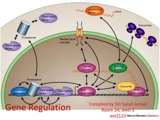
Factors Recruited to MHC II Promoters • Transcription factors that bind to conserved DNA elements: RFX (trimer of RFXANK, RFX5 and RFXAP) CREB (cAMP response element binding protein) NF-Y (trimer of A, B and C subunits) OCAB (“octamer” binding protein) • CIITA – MHCII Transactivator; acts as transcriptional “integrator” • BRG1 – Brahma-related gene 1; ATPase involved in remodeling nucleosome structure; vertebrate homolog of yeast SWI/SNF • CARM1 – Histone methylase • HAT – histone acetyltransferase; promotes “open” chromatin structure by acetylating core histones in nucleosomes • Factors in pre-initiation complex (PIC): p-TEFb, TAFs, TBP, etc. • RNAP II – RNA polymerase II; binds to Initiator element in MHC II promoter and catalyzes transcription elongation NOTE: All of these proteins except CIITA are ubiquitously expressed
CIITA is a member of the CATERPILLAR family of intracellular pattern recognition receptors Nucleotide-binding domain Leucine-rich repeats Transactivation domain Ting et al., Nat. Rev. Immunol. 6:183, 2006
Model for regulation of subcellular distribution of CIITA NLS = nuclear localization signal NES = nuclear export signal Ravalet al., J. Immunol. 170:922, 2003
Constitutive “Professional” APCs B cells Dendritic cells Inducible by IFN-g Many cell types Macrophages Epithelial cells Fibroblasts Others Expression of CIITA Molecules
Transcriptional Integration by CIITA Wright & Ting, Trends Immunol. 27:405, 2006
Structure of the CIITA gene locus p1 pIII pIV Wright & Ting, Trends Immunol. 27:405, 2006
Chromatin Remodeling Zika and Ting, Curr. Opin. Immunol. 17:58, 2005
Role of CIITA in Chromatin Remodeling Zika and Ting, Curr. Opin. Immunol. 17:58, 2005
Bare Lymphocyte Syndrome (BLS) • Loss of constitutive and inducible expression of all MHC II genes • Results in severe combined immunodeficiency because of loss of antigen recognition by T cells • Mutations involve factors associated with MHC II transcription, NOT the MHC II genes themselves RFXANK RFX5 RFXAP CIITA
Electrophoretic Mobility Shift Assay (EMSA) • In vitro assay of DNA-protein interactions • Isolate protein extracts (nuclear or whole cell) from cultured cells or tissues following experimental treatment. • Radiolabel short fragment of DNA or oligodeoxynucleotide containing a transcription factor binding site. • Incubate labeled DNA with protein extract to allow protein-DNA binding. • Separate protein-bound from unbound DNA by nondenaturating gel electrophoresis, and detect DNA by autoradiography. Protein-bound DNA will be “shifted” to a slower mobility than unbound DNA. • Variation: add an antibody against a specific transcription factor to the protein-DNA mix. The complex of antibody-protein-DNA will be shifted to a slower mobility than protein-DNA alone (“supershift”)
Chromatin Immunoprecipitation (ChIP) • Used to measure binding of proteins to DNA in native chromatin • Add formaldehyde to living cells to form DNA/protein and protein/protein crosslinks • Lyse cells and sonicate chromatin to break it into fragments with an average length of 500-1000 bp • Immunoprecipitate chromatin fragments with antibody to protein of interest • Heat precipitated chromatin to reverse protein-DNA crosslinks and digest with RNAse A and proteinase K to purify DNA • Amplify immunoprecipitated DNA fragments by PCR using primers for promoter of interest • Variation: use real-time PCR (see next slide) for a more quantitative measure of immunoprecipitated DNA.
Reverse Transcriptase (RT)-PCR • Used to measure steady-state levels of individual mRNAs • Isolate total cellular RNA from cultured cells or tissues following experimental treatment • Prepare complementary DNA (cDNA) by incubating RNA with random primers and reverse transcriptase • Amplify transcript from gene of interest by PCR, using sequence-specific primers • “Real-time” PCR uses fluorescent probes to analyze the level of amplified cDNA at each PCR cycle, and is more quantitative than “end-point” PCR, where the final amplified sample is analyzed by gel electrophoresis. • For more information about real-time PCR, visit: http://www.appliedbiosystems.com/support/tutorials/pdf/rtpcr_vs_tradpcr.pdf
Fluorescence-Activated Cell Sorting (FACS) • Used to measure protein expression in intact cells • For example, expression of the MHC II protein HLA-DR on the cell surface • Intact cells are incubated with fluorescent-labeled antibodies • Can measure multiple proteins on the same cell if you use different colors of fluorescent labels • Cells are sorted by machine and analyzed individually • Data are expressed as histograms with number of cells on the Y-axis and fluorescence intensity on the X-axis Immunofluorescence Microscopy • Used to analyze intracellular localization of proteins • Similar to FACS, but cells are fixed and stained on aslide, then imaged with a standard or confocal fluorescence microscope
Immunoblot (Western Blot) • Used to measure total protein expression in cells or tissues • Cells or tissues are lysed in a denaturing buffer, and proteins are separated based on molecular weight by denaturing polyacrylamide gel electrophoresis (SDS-PAGE). Larger proteins will migrate more slowly in the gel. • Separated proteins are transferred from the gel to a thin membrane of nylon or nitrocellulose (hence the term “blot”). • Individual proteins bound to the membrane are visualized by specific antibodies labeled with an enzyme or fluorochrome. The intensity of the band is proportional to the level of the protein. • Variation: ectopically expressed proteins with an epitope tag (e.g., “Flag” or “Myc”) can be detected with an epitope-specific antibody.
Constitutive Promoter Protein coding region Expression Vectors • Can be introduced into cells as plasmid or virus vectors • Can be transcribed/translated in vitro or introduced into living cells by transfection • Can encode wild-type or mutant form of protein • Proteins can be “tagged” with extra sequences, such as a “Flag” or “Myc” epitope
MHC II or CIITA Promoter “Reporter” protein Transcription Reporter Plasmids • Introduced into living cells by transfection • Activity of reporter protein (e.g., luciferase, CAT) measured as an index of transcriptional activity • Can be used to measure basal or inducible transcription • Can encode wild-type or mutant form of promoter
Strong Promoter GAL4-binding motif CIITA (WT or mutant) Weak Promoter Luciferase GAL4 GAL4 GAL4 GAL4 GAL4 CIITA Transactivation assay • A fusion protein is constructed by inserting a GAL4-binding motif at the 5’ end of the CIITA coding sequence. • A GAL4-dependent luciferase reporter plasmid is constructed by inserting 5 GAL4 sites upstream of a weak promoter driving luciferase transcription. • The GLA4:CIITA expression plasmid and GAL4:luciferase reporter plasmid are co-transfected into living cells. • Inside the nucleus, the GAL4-binding motif of the fusion protein binds to the GAL4 sites on the reporter plasmid. • The CIITA portion of the fusion protein transactivates the weak promoter. • Luciferase activity is measured as an index of transcriptional activity