Download
1 / 34
440 likes | 876 Views
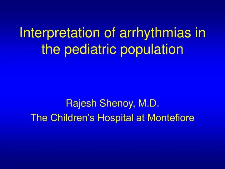
Interpretation of arrhythmias in the pediatric population. Rajesh Shenoy, M.D. The Children’s Hospital at Montefiore. Pediatric ECG Standardization. Paper speed is 25 mm/sec 1 large square = 0.2 sec 1 small square = 0.04 sec Voltage 10 mm/mv. Algorithm for reading ECGs.

E N D
Interpretation of arrhythmias in the pediatric population Rajesh Shenoy, M.D. The Children’s Hospital at Montefiore
Pediatric ECG Standardization Paper speed is 25 mm/sec 1 large square = 0.2 sec 1 small square = 0.04 sec Voltage 10 mm/mv
Algorithm for reading ECGs • Rhythm : Sinus or Non-sinus • Rate: 1500/ R-R interval (mm) • Axis: • QRS axis • T axis • Waves: • P wave (Atrial depolarization) • QRS complex (Ventricular depolarization) • ST segment • T wave (Ventricular depolarization) • U wave (Late phase of ventricular depolarization) • Intervals: • PR • QRS • QT/QTc
Rate • Calculate both the ventricular and atrial rate • In sinus rhythm, both are the same • Many methods • Heart rate scale • Adults: 300/(No. of large squares b/w R-R) • Children:1500/(No. of small squares b/w R-R)
Pediatric ECG – QRS Axis Hexaxial Reference System
Pediatric ECG – Determining Axis Using the Hexaxial Reference System
Normal axes P wave axis: In sinus rhythm, 0 to +90 QRS axis: 1st month +30 to +180 1-3 months +10 to +125 3 mo-3 years +10 to +110 > 3yrs +20 to +120 Adults -30 to +105 T wave axis: Positive in I and aVF
Pediatric ECG – Whats the Axis? What is the QRS Axis in this ECG?
Pediatric ECG – P Wave • Tall P waves ( > 3 mm) indicative of right atrial enlargement (P pulmonale) • Wide P waves ( > 0.10 sec) indicative of left atrial enlargement (P mitrale)
Pediatric ECG – PR Interval • Beginning of the P wave to beginning of QRS complex • Normal PR interval 0 – 1 mo 0.12 1 – 6 mo 0.14 6 – 12 mo 0.14 1 – 3 yr 0.15 3 – 8 yr 0.17 8-12 yr 0.18 12 – 16 yr 0.19 Adult 0.21 • Prolonged PR • Myocarditis • Rheumatic Fever • Digitalis toxicity • Hyperkalemia • Short PR • Preexcitation • Pompe disease
Pediatric ECG – QRS Complex • Q wave is narrow (0.02 sec) and short (5 mv) • Deep Q wave in left precordial leads = LVH • Q waves present in right precordial leads = RVH
QRS Duration Normal values < 3 yrs 0.07 s 3 – 8 yrs 0.08 s 8 – 12 yrs 0.09 s > 12 yrs 0.10 s Prolonged QRS Bundle branch block WPW syndrome
R and S waves • R/S progression: In older children, adolescents and adults – the R voltage increases, S voltage decreases • Voltage: Useful in determining hypertrophy of ventricles
Criteria for ventricular hypertrophy • RVH Tall R in V1 or deep S in V6 Upright T wave in V1 after 48-72 hrs Deep q wave in V1 RAD or strain pattern of T wave • LVH Tall R in V1 or deep S in V6 Deep q wave in V6 LAD or strain pattern of T wave
Pediatric ECG – ST segment • Normally isoelectric • Depression/Elevation of 1 mm in limb leads and 2 mm in precordial leads normal • Abnormalities • Pericarditis • Myocardial infarction • Digitalis
Pediatric ECG – T wave • Tall peaked T waves • Hyperkalemia • LVH • Flat low T waves • Normal newborns • Hypokalemia • Digitalis • Pericarditis • Myocarditis • Hypothyroidism • Inverted T wave in V1 normal in children
QTc interval Select the longest QT interval Measure QT interval from beginning of Q wave to end of T wave Measure R-R interval from the complex in which QT is measured to preceding R QTc interval (s)= QT interval (s) R-R interval(s)
QTc interval • Normal QTc interval varies depending on age • QTc prolonged >460 msec for male >450 msec for female • Causes of QTc prolongation • Prolonged QT syndrome • Macrolide antibiotics • Antifungals
Algorithm for interpretation of arrhythmias R-R interval Regular Irregular Continuously Intermittently Irregular Irregular QRS premature Or delayed? P-P regular? QRS rate? QRS duration P-wave morphology, P-wave mean vector, P-QRS
R-R interval Regular QRS Rate Decreased Normal Increased QRS Duration Normal Normal Normal P-wave morphology, P-wave mean vector, P-QRS relationship Normal Normal Normal Sinus bradycardia Normal sinusrhythm Sinus tachycardia
R-R interval Regular QRS Rate Normal QRS Duration Prolonged P-wave morphology, P-wave mean vector, P-QRS relationship Normal Bundle branch block W-P-W morphology
RBBB LBBB WPW
R-R interval Regular QRS Rate Normal QRS Duration Normal P-wave morphology, P-wave mean vector, P-QRS relationship Abnormal Atrial flutter Atrial fibrillation
Atrial flutter Atrial fibrillation
R-R interval Regular QRS Rate Normal QRS Duration Normal P-wave morphology, P-wave mean vector, P-QRS relationship Abnormal Left atrial rhythm (high or low) Low right atrial rhythm
Normal sinus rhythm High left atrial rhythm Low left atrial rhythm Low right atrial rhythm Junctional rhythm, retrograde ‘P’
R-R interval Regular QRS Rate Normal QRS Duration Normal P-wave morphology, P-wave mean vector, P-QRS relationship Abnormal Junctional rhythm Mobitz type II block Complete heart block
Fixed type II 2O AV Block 3O AV Block (AV dissociation)
R-R interval Irregular Continuously irregular P-P interval Regular Irregular Sinus arrhythmia Wandering atrial pacemaker 2O AV block, type I 2O AV block, type II
Sinus arrhythmia Type I 2O AV Block (Wenckebach) Type II 2O AV Block
R-R interval Irregular Intermittently irregular QRS premature QRS duration Normal Increased Conducted PAC Premature junctional beat PAC with aberration PVC
Premature supraventricular beat, conducted Premature junctional beat Premature ventricular beat
R-R interval Irregular Intermittently irregular QRS delayed QRS duration Normal Increased Type II AV block Non-conducted PAC Sinus pause+atrial escape Sinus pause+junct escape Sinus pause+vent escape
PAC, non-conducted Sinus pause, atrial escape Sinus pause, junctional escape Sinus pause, ventricular escape