Download
1 / 1
10 likes | 275 Views
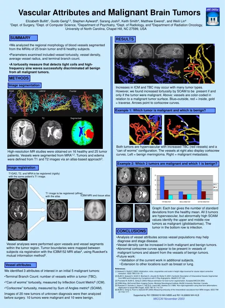
Axial. Lateral. Frontal. T1 T2. Segmented. T1. T2. T2. T1. T1. T2. Vascular Attributes and Malignant Brain Tumors. UNC CASILab. Elizabeth Bullitt 1 , Guido Gerig 2,3 , Stephen Aylward 4 , Sarang Joshi 5 , Keith Smith 4 , Matthew Ewend 1 , and Weili Lin 4

E N D
Axial Lateral Frontal T1 T2 Segmented T1 T2 T2 T1 T1 T2 Vascular Attributes and Malignant Brain Tumors UNC CASILab Elizabeth Bullitt1, Guido Gerig2,3, Stephen Aylward4, Sarang Joshi5, Keith Smith4, Matthew Ewend1, and Weili Lin4 1Dept. of Surgery, 2Dept. of Computer Science, 3Department of Psychiatry, 4Dept. of Radiology, and 5Department of Radiation Oncology, University of North Carolina, Chapel Hill, NC 27599, USA SUMMARY RESULTS • We analyzed the regional morphology of blood vessels segmented from the MRAs of 25 brain tumor and16 healthy subjects. • Parameters examined included vessel tortuosity, vessel density, average vessel radius, and terminal branch count. • A tortuosity measure that detects tight coils and high-frequency sine waves successfully discriminated all benign from all malignant tumors. METHODS Image segmentation Increases in ICM and TBC may occur with many tumor types. However, we found increased tortuosity by SOAM to be present if and only if the tumor were malignant. Above: vessels are color-coded in relation to a malignant tumor surface. Blue=outside, red = inside, gold = traverse. Arrows point to corkscrew curves. Example 1: Which tumor is malignant and which is benign? Both tumors are hypervascular with increased TBC (red vessels) and a “can of worms” configuration. The vessels at right also display corkscrew curves. Left = benign meningioma. Right = malignant metastasis. High-resolution MR studies were obtained on 16 healthy and 25 tumor patients. Vessels were segmented from MRA1,2. Tumors and edema were defined from T1 and T2 images via an atlas-based approach3. Example 2: Which 2 tumors are malignant and which 1 is benign? Image registration T1GAD, T2, and MRA to be registered (rigidly) with the same patient’s T1 image. T1 image to be registered (affine) with the atlas ICBM MRI and tissue atlas Graph: Each bar gives the number of standard deviations from the healthy mean. All 3 tumors are hypervascular, but abnormally high SOAM values identify the upper and middle row tumors as malignant (glioblastomas). The tumor in the bottom row is infection. CONCLUSIONS • Analysis of vessel attributes across vessel populations may help diagnose and stage disease. • Vessel density can be increased in both malignant and benign tumors. • Abnormal corkscrew curves appear to be present in vessels of malignant tumors and absent from the vessels of benign tumors. • Future work: • Validation of the current work in additional subjects. • Extension to other locations such as breast or lung. Vessel analyses were performed upon vessels and vessel segments within the tumor region. Tumor boundaries were mapped between subjects via registration with the ICBM152 MRI atlas4, using Rueckert’s mutual information method5. Vessel attributes References: [1] Aylward S, Bullitt E (2002) Initialization, noise, singularities and scale in height ridge traversal for tubular object centerline extraction. IEEE-TMI 21:61-75. [2] Bullitt E, Aylward S, Smith K, Mukherji S, Jiroutek M, Muller K (2001) Symbolic Description of Intracerebral Vessels Segmented from MRA and Evaluation by Comparison with X-Ray Angiograms. MedIA 5:157-169. [3] Prastawa M, Bullitt E, Gerig G (2003) Robust estimation for brain tumor segmentation. Accepted MICCAI 2003. [4] ICBM Atlas, McConnell Brain Imaging Centre, Montréal Neurological Institute, McGill University, Montréal, Canada. [5] Rueckert D, Sonoda LI, Hayes C, Hill DLG, Leach MO, Hawkes DJ (1999) Non-rigid registration using free-form deformations: Application to breast MR images. IEEE-TMI 18: 712-721 [6] Bullitt E, Gerig G, Pizer S, Aylward SR (2003) Measuring tortuosity of the intracerebral vasculature from MRA images. IEEE-TMI 22:1163-1171 • We identified 3 attributes of interest in an initial 5 malignant tumors: • Terminal Branch Count: number of vessels within a tumor (TBC). • “Can of worms” tortuosity, measured by Inflection Count Metric6 (ICM). • “Corkscrew” tortuosity, measured by Sum of Angles metric6 (SOAM). Images of 20 new tumors of unknown diagnosis were then analyzed before surgery. 10 tumors were malignant and 10 were benign. Supported by R01 EB000219 NIH-NIBIB and R01 HL69808 NIH-HLB MICCAI November 2003