Download
1 / 12
160 likes | 414 Views
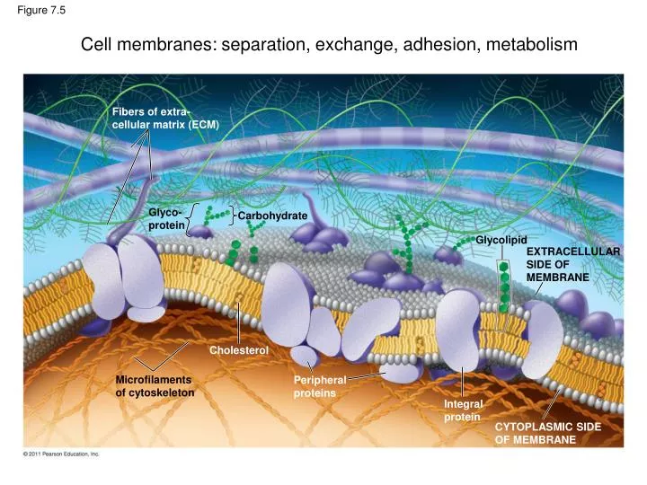
Cell membranes: separation, exchange, adhesion, metabolism. Fibers of extra- cellular matrix (ECM). Figure 7.5. Glyco- protein. Carbohydrate. Glycolipid. EXTRACELLULAR SIDE OF MEMBRANE. Cholesterol. Microfilaments of cytoskeleton. Peripheral proteins. Integral protein.

E N D
Cell membranes: separation, exchange, adhesion, metabolism Fibers of extra-cellular matrix (ECM) Figure 7.5 Glyco-protein Carbohydrate Glycolipid EXTRACELLULARSIDE OFMEMBRANE Cholesterol Microfilamentsof cytoskeleton Peripheralproteins Integralprotein CYTOPLASMIC SIDEOF MEMBRANE
(a) TEM of a plasmamembrane Outside of cell The basic structure of cell membranes Figure 6.6 Inside of cell 0.1 m Carbohydrate side chains Hydrophilicregion Hydrophobicregion Hydrophilicregion Phospholipid Proteins (b) Structure of the plasma membrane
TECHNIQUE Extracellularlayer Proteins Figure 7.4 Knife Plasma membrane Cytoplasmic layer RESULTS Inside of cytoplasmic layer Inside of extracellular layer
The phospholipid bilayer forms the structural basis of all cell membranes WATER Hydrophilichead Figure 7.2 Hydrophobictail WATER
Structure and amphipathic nature of membrane lipids Polar headgroup Non polar tails
Glycerophospholipid, ester type (fatty acids), found in bacteria and eukaryotes Plant, soybean C18:0, C18:2 Sphingomyelin, e.g. Brain, ester type (fatty acid and sphinganine) (2S,3R,4E)-2-acylaminooctadec-4-ene-3-hydroxy-1-Phosphocholine Glycerophospholipid; ether type (long chain alcohol) e.g. Mycoplasma fermentans 1,2-Di-O-Octadecenyl-sn-Glycero-3-Phosphocholine C18:1, C18:1 (ether) Archaeol; Glycerophospholipid, ether type (phytanol chains), found in archaea 1,2-Di-O-Phytanyl-sn-Glycero-3-Phosphocholine Hydrocarbon chain variability in phosphatidylcholines Glycerophospholipid (Plasmalogen), mixed ether and ester (fatty acid and long chain alcohol), found in brain, platelets
Membranes are dynamic structures Phospholipid bilayers are liquid crystalline: they are two dimensional liquids allowing lipid to move laterally along the membrane surface, but nor from monolayer to monolayer (flip-flop) Figure 7.6 Flip-flopping across the membraneis rare ( once per month). Lateral movement occurs107 times per second.
Viscous Fluid Figure 7.8 Unsaturated hydrocarbontails Saturated hydrocarbon tails (a) Unsaturated versus saturated hydrocarbon tails (b) Cholesterol within the animal cell membrane Desaturation of fatty acids produces fluidity Cholesterol
Snapshot of a molecular dynamics simulation of a lipid bilayer surrounded by water. Fig. 9.17 Voet, Fundamentals, 3rd
Membrane proteins and their association with the lipid bilayer Surface-bound Monotopic Integral, intrinsic Surface bound Peripheral, extrinsic Transmembrane Bi- and Polytopic Integral, intrinsic Surface-bound Lipid-anchored Integral, intrinsic
Figure 3.15 Hydrophobic surfaces in transmembrane proteins Hemoglobin Homo sapiens (1HGA) Top view Side view Polar (out) Non-polar Polar (in) Connexin 26 Homo sapiens (2ZW3) Reaction Center R.viridis (1PRC) Osmoporin OmpC E.Coli (1MPO)
Figure 3.18 Oligomerization and clustering of membrane proteins