Download
1 / 15
160 likes | 314 Views
Brain Computer Interfaces. Dr. Szczepan Paszkiel Department of Electrical, Control & Computer Engineering Institute of Control & Computer Engineering Opole University of Technology. Introduction – the biological aspect.

E N D
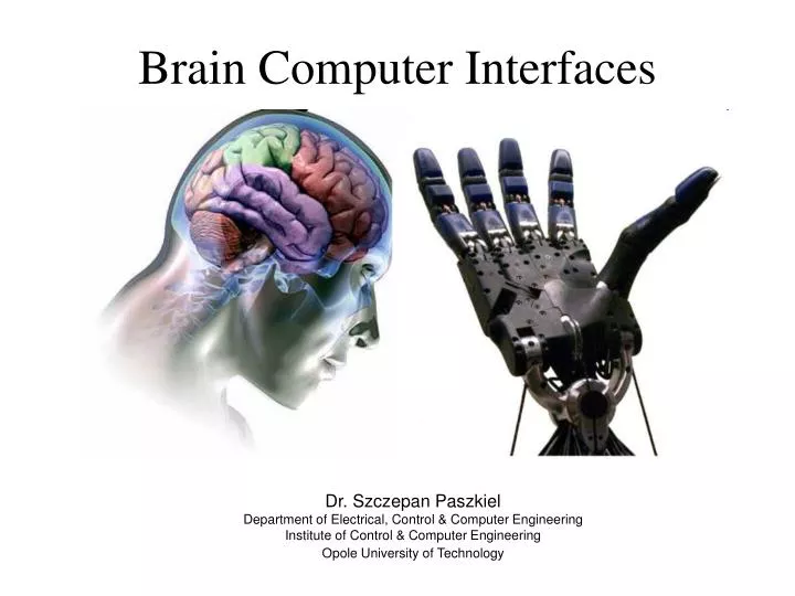
Brain Computer Interfaces Dr. Szczepan Paszkiel Department of Electrical, Control & Computer Engineering Institute of Control & Computer Engineering Opole University of Technology
Introduction – the biological aspect • There are mutual correlations between neurons in the human brain. We could identifytwo fractions of nerve cells - pyramidal cells and interneurons. • The cerebral cortex consists of a big number of neuron networks created by neurons that are close to each other. • Disruptive artefacts are crucial: • - biological: blood pressure, pulsation, nervous tics, etc. • - technical: electric system, medical equipment, etc.
Introduction - neurons The most essential part of the human brain is a neuron and it consists of:
A general diagram the non-invasive method of measurements There are two main methods to measure the electroencephalographic signal and these methods are a non-invasive and an invasive method. It is not possible to apply the invasive method because of law regulations and technical conditions. That is why research is conducted on the basis of the non-invasive method. EEG signal Electroencefalograph The non-invasive method depends on an acquisition of the EEG signal by means of active electrodes that are placed on the head of the subject.
System 10-20 Location of electrodes 10-20 System was elaborated by the International Federation of Clinical Neurophysiology • Types of electrodes: • mushroom-shaped • bowl-shaped There are two types of electrodes that are usedduring the EEG measurement. These are mushroom-shaped and bowl-shaped electrodes.
A device for research on the EEG signal - Emotiv Emotiv is one of the first producers of Brain Computer Interfaces in the world. The main window of the application Electrodes are placed on a frame and they communicate with a computer by means of Bluetooth. Equipment used in a Laboratory of biomedical measurements at Opole University of Technology. Green points mean that electrodes adjoin to the human skin in an adequate way. By means of them we could control software in the work station.
RestingPotential • There are two values of potentials in the nerve cells. Even though, the diffusion of the ions through the membrane is small during rest, after a long time it may cause a disappearance of essential differences in the concentration of the ions between an internal and external fluid and to a disappearance of the membrane potential as well. • A rest potential occurs when the value of the potential inside a neuron is around –70mV. • The occurrence of the rest potential is caused by the flow of ions of potassium according to a gradient of a concentration of the ions from inside to outside of a cell membrane.
Action Potential • The action potential can appear in every part of the neuron but the place of its regeneration is most often the initial part of an axon. • Every supraliminal stimulus causes the occurence of an action potential AP. The value of the potential inside a neuron is about +30mV. • Between the start of the stimulus and the start of the action potential is a small delay – latency. During the action potential neurons happen to be non-excitable and during a hyperpolarized consequent potential their excitability is lower. • These occurrences are described as an absolute and relative refraction. These refractions constitute a limitation for a maximum frequency which a neuron can make action potentials with.
Resting Potential - Action Potential There is a depolarization when the value of amplitude increases. When it decreases suddenly, there is a repolarization.
Neurotransmitters • Taking into account an effect made by a neuromediator, synapses may be divided into: • excitatory – a neurotransmitter (acetylocholine, noradrenaline, serotonin, dopamine) causes an opening of sodium canals and an inflow of soda to a cell and it leads to depolarization of postsynaptive membrane and the occurrence of postsynaptive excitatory potential (EPSP) • inhibitory - (GABA, glycine, somatostatine, alanine, prostoglandines) causes an opening of potassium and chlorine canals, a leak of potassium and an inflow of chlorine to the cell cause hyperpolarization of postsynaptive membranes which is the occurrence of inhibitory postsynaptive potential (IPSP)
Neurotransmitters Neuron In this slide we could see a visualization of a connection between neurons through a synapse. Synapse Neurotransmitter
EEG – twostages: excitatory - dormant The vertical column on the left side shows symbols of electrodes in the 10-20 standard.
1st - 8.01.2013 2nd - 22.01.2013