Download

1 / 42
460 likes | 513 Views
Immunodiagnosis of allergic diseases. Magdalena Żbikowska-Gotz. The diagnosis of allergy is based on: patient’s history subjective test skin prick test (SPT) measurement tIgE , asIgE mediators of allergic reaction. We can diagnose allergy: 1. in vivo 2. in vitro.

E N D
Immunodiagnosis of allergic diseases Magdalena Żbikowska-Gotz
Thediagnosis of allergyisbased on: • patient’shistory • subjective test • skin prick test (SPT) • measurementtIgE, asIgE • mediators of allergicreaction
We can diagnose allergy: 1. in vivo2. in vitro • 1. In vivo test based oninducing in vivo reaction to prove presence of antigen specific IgE: - in the skin (skin prick test) SPT - in organs (provocation tests – nasal, bronchial, conjunctival, digestal) provocation tests have very high value Caution!Risk factorfor systemic reaction and complications, tests may only be performed in hospital
2. In vitro testsAnalysis of the material collected from patient Material to analysis: blood, secretion products, sputum, tears, urine, NAL, BAL ( nasal lavage, bronchoalveolar lavage) Assays include: • estimating total IgE level in the blood serum • estimating specific IgE in the blood serum • tests evaluating the activity of cells in vitro -cells are extracted and induced specific allergens • tests evaluating the activity of cells in vivo - by mesuring levels ofmediators l which have been released in body
Advantages of application of in vitro tests • usage of high standardized allergens • estimation the level of parameters quantitatively • high specificity, sensitivity, repetitivity • objective results • safety (conducted outside body) • receive more information about the mechanism of disease • opportunity to estimate the severity of disease
Indications to in vitro methodsaccording to Clin. Rev. in Allergy Thismethodshould be applied: • in infants, small children and older patients with decreased reactivity or hyperactivity of skin due to different reasons. • in patients with intensive dermographism or extensive skin problems. • in patients unable to stop receiving medication influencing skin reactivity for the time required to do the test (depending on thedrug,onawerage 2 weeks) • in patients with history of systemic reaction during skin tests. • when diagnosing of hymenoptera allergy (skin tests dangerous). • whentherearedifference between skin tests and clinicalhistory
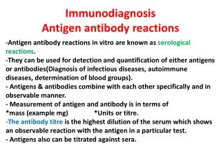
Immunological diagnostic – in vitro test • they are based on the affinity of antigen and antibody, and creating complex on theabsorption surface • stability of complex influences the quality of methods (specificity, sensitivity) sensitivity of the method: - defined as the smallest number of examinated substance, which can be measured (difference from zero) - depends of the marker used on the marked antibodies or antigens (radioisotopes, fluorescents markers, enzymatic markers, luminescent markers)
Precision of method measure of precision is the difference between the value from measurement and the actual value it depends of: - immunoabsorbing surface (the size of surface) - immunoabsorbing surface (microplates, magnetic spheres, blotting paper discs, microparticle, liquid phase – large surface of absorption) Specificy of methods method is specific if it determines only one substance in the examinated material the affinity of Ag-Ab complex depends on used monoclonal antibodies (revealing high specificity)
Test tIgE – total IgE measures the quantitative amount of total IgE in serum or plasma IgE anibodies are produced by the body as a result of repeated allergenic stimulation Test as IgE - allergen specific IgE specific IgE measures IgE antibodies to specific allergens in human serum or plasma specific IgE antibodies appear as a result of sensitization to an allergen and can be detected following exposure
Hypersensitivity type1 • Total IgE, indicate intensity of humoral reaction - level of IgE in the blood serum is correlated with age - level of IgE increases from birth (0-1 kU/l) to puberty, then it decreases to a constant level in the age of 20-30 years • normal level of IgE is oscilates around 40-70 kU/l • concentration of total IgE > 10 kU/l in the first year of life and total IgE > 100 kU/l in adults is considered incorrect • concentration < 30 kU/l allows usually to rule out atopy (exception are patients with hymenoptera allergy, medication – penicilin)
Total IgE level depends on: • presence of atopy • number of sensitizing allergens and strength of specific allergy • exposition time • range of multiorgan symptoms of allergy • Estimating total IgE has an „orientation” value because: • low level of total IgE does not rule out atopy (to note! - IgE is cytophile Ab which after being produced is bound by receptors on the effector cells’ surface),half-life IgE in blood is 2-3 days
Estimating total IgE has an „orientation” value because: • -low level of total IgE does not rule out atopy (to note! - IgE is cytophile Ab which after being produced is bound by receptors on the effector cells’ surface),half-life IgE in blood is 2-3 days - correct or higher levels of IgE can be also observed in the allergy manifestation -higher levels of IgE may be also observed in patients with other diseases than allergy (parasites, hiperimmunoglobulinemia IgE, immunosuppression, AIDS, burn victims, hepatitis, nephrotic syndrome, virus infections, mucowiscydose, smokers, psoriatic arthritis)
Analysis of relation between the level of total IgE and different diseases Ramosa, Allergy 2002, 57 • Healthy (av. 74,2 ± 51,3) • Food allergy (av. 83,3 ± 67,0) • Urticaria (av. 96,8 ± 72) • Allergic rhinities (av. 327,5 ± 137) • Atopic asthma (av. 411,6 ± 283) • higher level of total IgE is observed in over 60% of patients with allergy, usually connected with increasing of antigen specific IgE • similar levels may be the result of eliminating diet-lack allergic induce
Themeasurement of asIgEis the most important in vitro analysis. It: estimates allergy to specific allergen determines the level of sensitization hasresultswhich are quantitative and expressed in international units (IU/ml, kU/l), ranked in classes (0-VI) isusefulin patients with allergymostly to pollen, dust mite, food (when causing anaphylactic reaction), animal epidermis, fungal moulds and other food allergens.
diagnostic sensitivity and specificity of asIgE for inhalatory allergens is 90-94% • diagnostic sensitivity and specificity of asIgE for food allergens is lower (weeker for animal allergens, stronger for plant allergens) - diagnostic sensitivity and specificity of asIgE for hymenoptera allergens is high (above 80%)
How the concentration levels translate into different classes? To note! – presence of as IgE means sensitivity but does not equal allergy Allergy = presence of IgE and clinical symptoms
Other tests: • Screening tests, whichcanrevealasIgEagainstbasicallergensencompass: • Panels (inhalatoryor food) • Phadiatops (inhalatoryor food) • Pediatrics phadiatop(inhalatory and food) • Fast screen Requiredare: -at least one important allergen of each group (inhalatory, food) intests low sensitivity maylead to diagnostic mistakes resultsaredisplayed as neg. or pos.
Phadiatop,Panel,Phadiatop Infant -are tests that differentiate atopic and non- atopic diseases -may be the first step taken when testing for atopic allergy -the test comprise a balanced mixture of relevant allergens that react with specific IgE anibodies -tests demonstrate the presence of atopic allergy to common allergens relevant for adults and small children
To evaluate the level of total IgE, specific IgE and screening tests we use the following methods of immunodetection: 1. RIA (radioimmunoassay) 2. EIA (enzymeimmunoassay) 3. FEIA (fluoroenzymoimmunoassay)
To acknowledged and verified examinations in the diagnosis of allergy belong also: • tests estimating the ability to activate cells in vitro • tests estimating activity of cells in vivo by measuring mediators 1&2 applied less frequently in routine cases but mainly in complex ones We estimate specific markers of cells involved in the patomechanism when atopicclinical history is accompanied by: • deficiency clinical symptoms or • negative skin prick tests or • normal level of total and specific IgE.
Tests of cell activity in vitro • BAT : evaluating the expression of specific basophil activation surface markers (CD 63 and CD 203c). -difference CD 203c and CD 63 CD63: directly linked to basophil degranulation upon allergen contact only expressed upon allergen contact CD203c: upregulation upon allergen contact
BASO-CAST • estimating the activation of basophils through stimulation of specific sensitizing factor • at the same time the test estimates: • activation marker BA CD63 with cytometric method • in supernants we estimate the level of LTC4 immunoenzymatic - test is useful in estimating the hypersensitivity to pollen, hymenoptera venom, latex, foods, aspirin • sensitivity and specificy of test is bigger then specific IgE test Flow cytometry is a method of estimating physical andbiological properties of each cell in the moment oftheir suspension flow through the detector.
CAST-ELISA test (Cellular Antigen Stimulation Test) - estimatesthe concentration of leukotriens (depending of the level of cell activation) - draws on isolating leucocytes (in presence of IL3 and IL5, and specific allergens) releasing sulfidoleukotriens, that are next marked using immunoenzymatic method (Abs anty LTC4, LTD4, LTE4) - indicates high level of correlation between skin prick tests and specific IgE in the blood serum - recommended: food allergy, antibiotics, hymenoptera venom
Test of releasing histamine from leucocytes or mast cells after enzymatic dispersion of tissue: • estimates histamine concentration – early mediator of specific antigen-sIgEreaction • is helpful in determinating sensitizing factor • draws on methods: enzymatic, micromethod HR-MM (histamine release microfiber method), affinity of histamine to fiber glass • has little clinical value
Examination of cellactivity- in vivo - Tests estimating concentration of mediators created in the biological fluids such as:blood, tears, urine and others during allergic reaction Tryptase (tetrameric endoprotease) • specific indicator of degranulation (activation) of mastocytes – lack of Ne, Eo, L, M • mature beta-tryptase stored in granula is released during mast cell activation • indicator of early phase allergic reaction (preform marker) • the transiently increased levels of tryptase serve as a clinical marker confirming severe reactions as anaphylaxis. • evaluation using RIA, FEIA method • there is no established connection between concentration of tryptase in blood serum and severity of asthma
Eosinophils • role in development and sustaining the late phase of allergic reaction • contain granulations that store protein substances (MBP, ECP, EPX/EPN, EP0) • 2 types of eosinophils cells – hypo- and normodensity • level in the blood or tissues can be a measure of the strength of allergic inflammation interpretation of the result should be careful because eosinophils take part in many diseases – parasites, infectious diseases, gastritis, sarcoidosis.
ECP (Eosinophil cationic protein) • main mediator of eosinophiles • exponent of eosinophils activation (neutrophilia?) • material – blood serum, tears • high concentration ECP in the blood serum of patients with exacerbation of asthma, allergic rhinitis, atopic dermatitis, other nonallergic diseases • ECP as a factor does not difference the place, where the allergic inflammation is present
Test ECP ECPmeasures the level of Eosinophil Cationic Protein released in serum ECP is a highly cytotoxic protein found in eosinophil granules when eosinophils in the airway are activated ,they undergo degranulation causing airway epithelial damage and desquamation ECP is released from the activated eosinophils during the inflammation process
so far diagnostic methods have drawn on allergen extracts which identified source of allergy • today it is possible to diagnose allergy by using the components of allergens UniCap, ISAC Using components is prerequisite for detecting cross-reactivity in allergy
Allergens consist of components Components consist of epitopes Different allergens have homological epitopes or epitopes with high level of similarity
Cross allergy -reasons of cross reactivity- • there is a restricted number of elements that build epitops • sometimes identical or similar epitops occur in different, unrelated allergens • therefore specific antibody against one antigen can cross react with another allergen than the one that induced the immunological response • typically cross reactivity is IgE dependent • when patient suffers both from inhalatory and food allergy the mechanism can be either co-reactivity or cross reactivity • describing mechanism of cross reactivity is possible partly because panallergens (commonplace allergens) were discovered • panallergens include– profilins, carbohydrate determinants – CCD and lipid transporting proteins – LTP, serum albumin, TLP, tropomiosin, storage protein, PR10.
Cross reactivity happens when AbIgE produced originally against one allergen (commponent) recognises and binds to similar component from another source (allergen)
Diagnosis of classical cross reactivity is based on tests using recombinated components of allergens isolated by molecular biology method such as ImmunoCAP ISAC
ImmunoCAP ISAC IgE • solid-phase immunoassay to detect asIgE • semiquantitative test in vitro estimate concentration Ab in serum to 112 allergens molecules (components) from >50 different allergens sources • allergen components that are immobilized on a solid substrate in a microarray format are incubated with human serum • binding of the specific IgE antibodies to the immobilized allergen components is detected by addition secondary flurescence labeled anti-human IgE antibody
ImmunoCAP ISAC IgE • the procedure is followed by image aquisition using appropiate microarray scaner • the level of concentration asIgE is proportionality to intensity of determinate fluorescence • result of the research is analyzed with propertary software and expressed in standarized units (ISU - E)
BENEFITS of using ImmunoCAP ISAC 1. concurrent analysis of different biological interactions - which is important for the suspected allergy to a variety of factors 2. allows to obtain a result of a large number of allergen components, including the formation of allergic profile of the patient 3. allows to explain the cross-reactivity that occurs between allergens
BENEFITS of using ImmunoCAP ISAC 4. can point to other potential sources of allergies, especially when the sensitization image is unclear and direct cause of allergy is difficult to diagnose 5. allows to assess the risk of severe allergic reactions (assessed the presence of highly allergenic components) 6. allows for the correct classification of patients for specific immunotherapy SIT