Download
1 / 43
470 likes | 1.22k Views
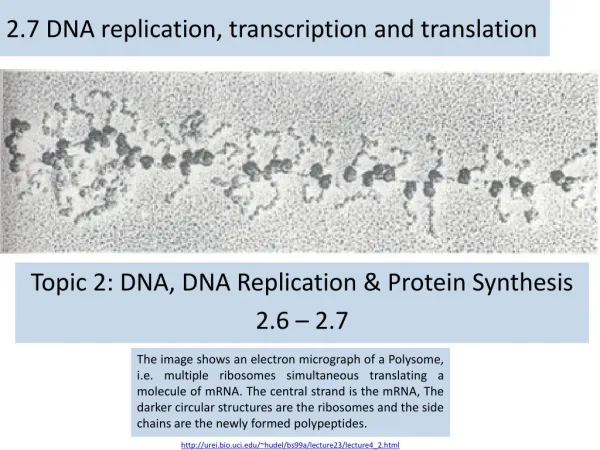
Chapter 25: Nucleotides, Nucleic Acids, and Heredity. DNA Replication, Transcription and Translation. DNA Replication, Transcription and Translation. Overview. DNA DNA mRNA Protein. Replication Transcription Translation. A. A. A. A. A. α.

E N D
Chapter 25: Nucleotides, Nucleic Acids, and Heredity DNA Replication, Transcription and Translation DNA Replication, Transcription and Translation
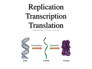
Overview DNA DNA mRNA Protein Replication Transcription Translation A A A A A α Francis Crick (1958): Central Dogma of Molecular Biology
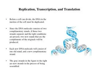
DNA Structure • Deoxyribonucleic acid (DNA) • Structure: Double Helix (Two strands) • Function: long-term storage of information
DNA Replication A A Enzymes: 1-Helicase 2-DNA Polymerase 3-Topoisomerase 4-DNA primase 5-DNA Ligase
RNA Structure • Ribonucleic acid (RNA) • Structure: Single strand • Functions: • Four bases : • (adenine, cytosine, guanine and uracil) • mRNA: information carrier • rRNA: Ribosomes Constituent • tRNA: amino acid transporter
Transcription A A
Protein Structure • Polypeptide: amino acids arranged in a linear chain • Structure: multiple linear and 3D structures • Functions: • Enzymes • Cell signaling (insulin) • Ligand binding (antibodies) • Transport • Structural
Translation α • Initiation:the small subunit of the ribosome binds to 5' end of the mRNA with the help of initiation factors • Elongation:additional amino acid is added to the growing polypeptide chain • Termination:one of the three termination codons moves into the A site A Video
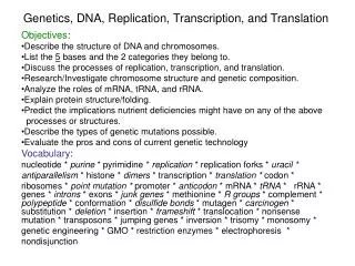
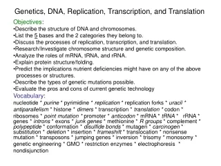
The Molecules of Heredity • Each cell of our bodies contains thousands of different proteins. • How do cells know which proteins to synthesize out of the extremely large number of possible amino acid sequences? • From the end of the 19th century, biologists suspected that the transmission of hereditary information took place in the nucleus, more specifically in structures called chromosomes. • The hereditary information was thought to reside in genes within the chromosomes. • Chemical analysis of nuclei showed chromosomes are made up largely of proteins called histones and nucleic acids.
The Molecules of Heredity • By the 1940s, it became clear that deoxyribonucleic acids (DNA) carry the hereditary information. • Other work in the 1940s demonstrated that each gene controls the manufacture of one protein. • Thus the expression of a gene in terms of an enzyme protein led to the study of protein synthesis and its control.
Nucleic Acids There are two kinds of nucleic acids in cells: • Ribonucleic acids (RNA). • Deoxyribonucleic acids (DNA). Both RNA and DNA are polymers built from monomers called nucleotides. A nucleotide is composed of: • A base, a monosaccharide, and a phosphate.
Nucleosides Nucleoside:A compound that consists of D-ribose or 2-deoxy-D-ribose bonded to a purine or pyrimidine base by a -N-glycosidic bond.
Nucleotides Nucleotide: A nucleoside in which a molecule of phosphoric acid is esterified with an -OH of the monosaccharide, most commonly either at the 3’ or the 5’-OH.
Nucleotides Adenosine 5’-triphosphate (ATP) serves as a common currency into which energy gained from food is converted and stored.
DNA—Primary (1°) Structure For nucleic acids, primary structure is the sequence of nucleotides, beginning with the nucleotide that has the free 5’ terminus. • The strand is read from the 5’end to the 3’end. • Thus, the sequence AGT means that adenine (A) is the base at the 5’ terminus and thymine (T) is the base at the 3’ terminus.
Structure of DNA and RNA Figure 25.2 Schematic diagram of a nucleic acid molecule. The four bases of each nucleic acid are arranged in various specific sequences. The base sequence is read from the 5’ end to the 3’ end.
DNA—2° Structure Secondary structure: The ordered arrangement of nucleic acid strands. • The double helix model of DNA 2° structure was proposed by James Watson and Francis Crick in 1953. Double helix: A type of 2° structure of DNA in which two polynucleotide strands are coiled around each other in a screw-like fashion.
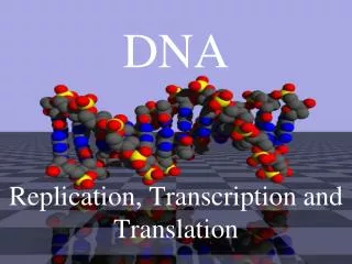
THE DNA Double Helix Figure 25.4 Three-dimensional structure of the DNA double helix.
Base Pairing Figure 25.5 A and T pair by forming two hydrogen bonds. G and C pair by forming three hydrogen bonds.
Superstructure of Chromosomes DNA is coiled around proteins called histones. • Histones are rich in the basic amino acids Lys and Arg, whose side chains have a positive charge. • The negatively-charged DNA molecules and positively-charged histones attract one another and form units called nucleosomes. Nucleosome: A core of eight histone molecules around which the DNA helix is wrapped. • Nucleosomes are further condensed into chromatin. • Chromatin fibers are organized into loops, and the loops into the bands that provide the superstructure of chromosomes.
Superstructure of Chromosomes • Figure 25.8
Superstructure of Chromosomes • Figure 25.8 cont’d
Superstructure of Chromosomes • Figure 25.8 cont’d
DNA and RNA The three differences in structure between DNA and RNA are: • DNA bases are A, G, C, and T; the RNA bases are A, G, C, and U. • the sugar in DNA is 2-deoxy-D-ribose; in RNA it is D-ribose. • DNA is always double stranded; there are several kinds of RNA, all of which are single-stranded.
RNA Table 25.3 The roles of Different kinds of RNA
Structure of tRNA Figure 2.10 Structure of tRNA.
Structure of rRNA • Figure 25.11 The structure of a typical prokaryotic ribosome.
Ribosome • Figure 25.11 cont’d
Genes, Exons, and Introns Gene: A segment of DNA that carries a base sequence that directs the synthesis of a particular protein, tRNA, or mRNA. • There are many genes in one DNA molecule. • In bacteria, the gene is continuous. • In higher organisms, the gene is discontinuous. Exon: A section of DNA that, when transcribed, codes for a protein or RNA. Intron: A section of DNA that does not code for anything functional.
Genes, Exons, and Introns • Figure 25.12 The properties of mRNA molecules in prokaryotes versus eukaryotic cells during transcription and translation.
Genes, Exons, and Introns • Figure 2.12 cont’d
Replication of DNA The DNA in the chromosomes carries out two functions: • (1) It reproduces itself. This process is called replication. • (2) It supplies the information necessary to make all the RNA and proteins in the body, including enzymes. Replication begins at a point in the DNA called the origin of replication or a replication fork.
Replication of DNA Figure 25.13 General features of the replication of DNA. The two strands of the DNA double helix are shown separating at the replication fork.
Replication of DNA The replication of DNA occurs in number of distinct steps. 1. Opening up of the superstructure of the chromosomes. One key step is this process is acetylation-deacetylation of lysine residues on histones. This reaction eliminates some of the positive charges on histones and weakens the strength of the DNA-histone interaction.
Replication of DNA 2. Relaxation of Higher-Order Structures of DNA. Tropoisomerases (also called gyrases) temporarily introduce either single-or double strand breaks in DNA. Once the supercoiling is relaxed, the broken strands are joined together and the tropoisomerase diffuses from the location of the replication fork. • Unwinding the DNA Double Helix. Replication of DNA molecules starts with the unwinding of the double helix which can occur at either end or in the middle. Special unwinding proteins called helicases, attach themselves to one DNA strand and cause the separation of the double helix.
Replication of DNA 4. Primers/Primases Primers are short—4 to 15 nucleotides long—RNA oligonucloetides synthesized from ribonucleoside triphosphates. They are needed to initiate the primase-catalyzed synthesis of both daughter strands. 5. DNA Polymerase Once the two strands are separated at the replication fork, the DNA nucleotides must be lined up. In the absence of DNA polymerases, this alignment is extremely slow. The enzyme enables complementary base pairing with high specificity. While bases are being hydrogen bonded to their partners, polymerases join the nucleotide backbones.
Replication of DNA Along the lagging strand 3’—>5”, the enzymes can synthesize only short fragments, because the only way they can work is from 5’ to 3’. These resulting short fragments consist of about 200 nucleotides each, named Okazaki fragments after their discoverer. 6. Ligation The Okazaki fragments and any nicks remaining are eventually joined by DNA ligase.
DNA Repair The viability of cells depends on DNA repair enzymes that can detect, recognize, and remove mutations from DNA. The most common repair mechanism is called base excision repair (BER). This pathway contains two parts. 1. A specific DNA glycosylase recognizes the damaged base. It hydrolyzes the N-C’ -glycosidic bond between the damaged base and the deoxyribose, then releases the damaged base. The sugar-phosphate backbone is still intact. 2. The backbone is cleaved by a second enzyme, an endonuclease. A third enzyme, an exonuclease, then liberates the sugar-phosphate unit of the damaged site. 3. In the synthesis step, DNA polymerase inserts the correct nucleotide and the enzyme DNA ligase seals the backbone to compete the repair.
How Do We Amplify DNA? • To study DNA for basic and applied scientific purposes, we must have enough of it to work with. • Millions of copies of selected DNA fragments can be made within a few hours with high precision by a technique called polymerase chain reaction (PCR). • To use PCR, the sequence of a gene to be copied or at least a sequenced segment bordering the desired DNA must be known. • In such a case, two primers that are complementary to the ends of the gene or to the bordering DNA can be synthesized. The primers are polynucleotides consisting of 12 to 16 nucleotides. When added to the target DNA segment, they hybridize with the end of each strand of the gene.
How Do We Amplify DNA A polymerase extends the primers in each direction as individual nucleotides are assembled and connected on the template DNA. In this way two copies are created. The two-step process is repeated (cycle 2) when the primers are hybridized with new strands and the primers extended again. At this point, four new copies have been created. The process is continued, and in 25 cycles, 225 or some 33 million copies can be made. This process is practical because of the discovery of heat-resistant polymerases isolated from bacteria that live in hot thermal vents on the sea floor. A temperature of 95°C is required to unwind the double helix to hybridize the primer to the target DNA.
How Do We Amplify DNA? • Figure 25.16 Polymerase chain reaction (PCR). Oligonucleotides complementary to a given DNA sequence prime the synthesis of only that sequence.