Download
1 / 96
980 likes | 1.19k Views
Cell Division. Biology. Storing genetic information. Genetic information is stored as DNA (a type of nucleic acid ) Human DNA contains 6 billion nucleotides in each cell (sperm and egg have half that) Genes are segments of DNA that control hereditary traits

E N D
Cell Division Biology
Storing genetic information • Genetic information is stored as DNA (a type of nucleic acid) • Human DNA contains 6 billion nucleotides in each cell (sperm and egg have half that) • Genes are segments of DNA that control hereditary traits • They provide a code that tells the body which proteins to make • Normally, DNA in the nucleus is in long strands called chromatin. • During cell division, the chromatin is coiled into compact structures called chromosomes
Types of Chromosomes • Sex Chromosomes-determine the gender of an organism • In humans, XX = female, XY = male • All other chromosomes are autosomes • Each species has a characteristic number of chromosomes • Cat = 32 • Carrot = 18 • Adder’s tongue fern = 1,262 • Humans have 46 • 2 are sex chromosomes • 44 are autosomes
Homologous Chromosomes • Chromosomes are arranged in pairs-you have 23 pairs • For each pair, you received one from your mother and one from your father • These two chromosomes code for the same types of proteins, but may have different information on them • These two are called homologous chromosomes or homologues • They are the same size, shape, and carry genes for the same traits
Karyotype • In a karyotype, chromosomes are photographed, cut out, and arranged in pairs according to size, shape, and bands. • In other words, they are lined up with their homologue • A karyotype can detect certain abnormalities, such as Down Syndrome • The sex chromosomes are put at the end and the karyotype will show if the baby is male or female
Types of Cells • Diploid cells have a complete set of chromosomes-46 arranged in 23 pairs. It is sometimes abbreviated as 2n. • All cells except the reproductive cells (sperm, egg) are diploid. • Haploid cells have half a set; they have one of each of the 23 pairs-23 total chromosomes. Abbreviated 1n • The only cells that are haploid are the reproductive cells (sperm, egg) • Why are sex cells haploid?
Why do cells divide? • Cell division occurs the most in young, growing individuals (embryo, fetus) and when cells are damaged or destroyed. • Some cells divide throughout life (skin, lining of intestine) while some don’t divide once fully grown (nerve). • Why are injuries to the brain and spinal cord often permanent? • Cells divide rather than just getting bigger because the surface area to volume ratio becomes too small.
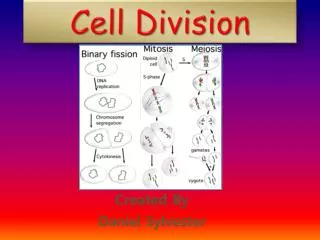
Cell Division in Prokaryotes • Called binary fission • DNA copies itself, cell grows, cell splits • Result: exact copy of parent (asexual)
Cell Division in Eukaryotes • Making an exact copy of a eukaryotic cell is called mitosis • Mitosis technically refers to the division of the nucleus; it is followed by cytokinesis, the division of the cytoplasm • Every cell in your body that divides undergoes mitosis (a different process makes sperm and egg) • Why would you want your skin cells to be an exact copy?
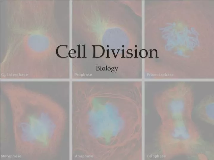
Interphase • Period in between cell division • “resting stage” • Most of a cell’s life is spent in interphase • G1: First gap: Cell grows • S: Synthesis of DNA-DNA is replicated • G2: Second Gap: Cell continues to grow, cell gets ready to divide
Mitosis: Prophase • Chromatin shortens and becomes chromosomes • Because DNA has already replicated, each chromosome has 2 identical parts • These identical parts are called “sister” chromatids and are held together at a point called the centromere • Note: They look like an X, but don’t confuse them with the X sex chromosomes
Mitosis: Prophase (cont.) • The nucleolus and nuclear envelope disappear (break down) • Dark spots called centrosomes appear. Inside, in animal cells, are T-shaped structures called centrioles • There are also short protein fibers called astral rays. • Together, astral rays + centrioles are called asters
Mitosis: Prophase (cont.) • As the centrosomes move toward opposite poles, spindle fibers, made of microtubules, radiate out. • There are two types of spindle fibers: • Kinetochore: from centromere to centriole (part way) • Polar: from centriole to centriole (full length)
Mitosis: Metaphase • Kinetochore fibers move the chromosomes to the center of the dividing cell • The chromosomes will be lined up along the equator
Mitosis: Anaphase • The chromatids of each chromosome pull apart and move (pulled by the centromere) toward opposite poles. • The chromosomes no longer look like Xs because they were pulled apart.
Mitosis: Telophase • Opposite of Prophase • Asters, spindle fibers disappear • Nuclear envelope, nucleolus reappear • Chromosomes return to chromatin
Cytokinesis • Division of the Cytoplasm • Animals: A cleavage furrow pinches in • Plants: A cell plate divides the cell
Cell Cycle • The events occur in motion, not distinct stages. • Most cells will repeat these stages, so the process is called the cell cycle • Result of Mitosis: 2 offspring cells, identical to the original cell (1 2)